Atharva Gautam - LGP-26-B2
 May 26, 2026 • 10:17 AM UTC
 India
India 340x Magnification
 Plants
Atharva Gautam
Learn about the author...
2posts
0comments
0locations
View in Media Gallery
Day 1:
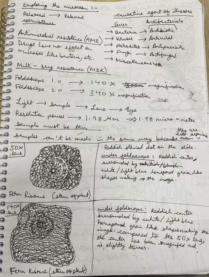
Observing the fern rhizome under the foldscope: The sample had already been prepared for us, with a red-stained dot on the glass slide. We learned how to properly utilise the foldscope. We could also see a lot of small details quite clearly. The reddish stain made some parts stand out, especially the small dotted structures and cells. Some areas looked darker red while others were lighter. I also observed tiny hexagonal and oval-shaped patterns that looked like dots spread across the section. We observed it at 50x, 140x, and 340x lenses.
Observing the fern rhizome under the foldscope: The sample had already been prepared for us, with a red-stained dot on the glass slide. We learned how to properly utilise the foldscope. We could also see a lot of small details quite clearly. The reddish stain made some parts stand out, especially the small dotted structures and cells. Some areas looked darker red while others were lighter. I also observed tiny hexagonal and oval-shaped patterns that looked like dots spread across the section. We observed it at 50x, 140x, and 340x lenses.

View in Media Gallery
We also observed the fern rhizome using the 340x lens:

View in Media Gallery
Day 2:
Observing onion cells under the foldscope: For this observation, I prepared the onion peel sample by myself. First, I carefully removed a thin transparent layer from the onion and placed it on a slide with cellotape. When I looked through the foldscope, I could clearly see tiny box-shaped onion cells arranged in neat rows. The cells looked like small bricks joined together. The cell walls were visible and formed clear boundaries around each cell. Some parts appeared lighter while others looked slightly darker. It was really interesting how organised the onion cells were. The onion peel under the foldscope showed a detailed microscopic structure. We observed it at 50x, 140x, and 340x lenses. Preparing the sample by myself was also slightly challenging, as it was my first time attempting such a task, as well as the added slippery nature of the onion peel. But I was able to eventually get the sample prepared on the slide.
140x lens:
Observing onion cells under the foldscope: For this observation, I prepared the onion peel sample by myself. First, I carefully removed a thin transparent layer from the onion and placed it on a slide with cellotape. When I looked through the foldscope, I could clearly see tiny box-shaped onion cells arranged in neat rows. The cells looked like small bricks joined together. The cell walls were visible and formed clear boundaries around each cell. Some parts appeared lighter while others looked slightly darker. It was really interesting how organised the onion cells were. The onion peel under the foldscope showed a detailed microscopic structure. We observed it at 50x, 140x, and 340x lenses. Preparing the sample by myself was also slightly challenging, as it was my first time attempting such a task, as well as the added slippery nature of the onion peel. But I was able to eventually get the sample prepared on the slide.
140x lens:

View in Media Gallery
Sign in to commentNobody has commented yet... Share your thoughts with the author and start the discussion!
 0 Applause
 0 Comments