Cells in an orange flower petal
 May 30, 2019 • 5:28 PM UTC
 United States
 140x Magnification
 Unknown
Sophie Li
Learn about the author...
9posts
0comments
2locations
View in Media Gallery
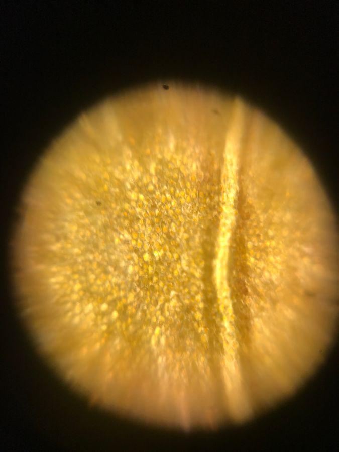
Location: This petal sample was taken from outside of Page house. Page house is a building on the campus of the California Institute of Technology in Pasadena, California.
Time: The sample was taken on May 30th at 16:13
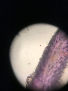
The sample was obtained by taking a petal from the flower pictured below. I cut a small section of a stuck it onto the sticky Foldscope slide.
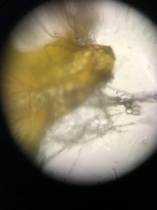
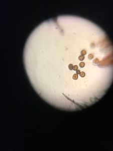
Description: In the image, we can see small circles. These are most likely the cells of the flower. The entire flower had an orange pigment, which can also be seen on this image. The line that runs through the image is a vein of the flower. Here the petal was thicker and had a lighter pigment. It is probably used to transport water and nutrients throughout the petal.
Time: The sample was taken on May 30th at 16:13
The sample was obtained by taking a petal from the flower pictured below. I cut a small section of a stuck it onto the sticky Foldscope slide.
Description: In the image, we can see small circles. These are most likely the cells of the flower. The entire flower had an orange pigment, which can also be seen on this image. The line that runs through the image is a vein of the flower. Here the petal was thicker and had a lighter pigment. It is probably used to transport water and nutrients throughout the petal.
View in Media Gallery
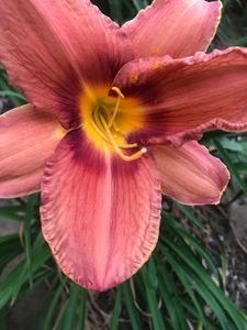
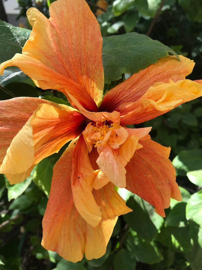
Here is an image of the flower from which the petal was taken. It was on a large bush growing on a wall.
#caltechbi1
#caltechbi1
Sign in to commentNobody has commented yet... Share your thoughts with the author and start the discussion!
 0 Applause
 0 Comments