LGP Day 1 - Exploring the microcosmic world
 Jun 05, 2024 • 9:36 AM UTC
 India
India 340x Magnification
 Workshops and Events
Navya
Learn about the author...
2posts
0comments
1locations

View in Media Gallery
Date: 3rd June 2024
Day: Monday
Week 3 batch
Time: 4:15pm
Venue: AC04-005
Name of student: Navya Agarwal
Day: Monday
Week 3 batch
Time: 4:15pm
Venue: AC04-005
Name of student: Navya Agarwal

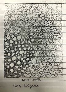
This is a picture and drawing of a Fern Rhizome. This is under the 140X magnification.
We can observe a very distinct layered structure. The inside consisted of loosely packed cells which were surrounded by a black filament. As we go outwards, the cells become more compactly packed while the cells become smaller.
The third layer has larger cells, however they are also very compactly packed. They are mostly rectangular in shape. The last two layers have smaller and very densely packed structures.
A blue tint has been added to this Fern Rhizome so that the cells and components in the cells are better and more clearly visible.
____________________________________________________________________


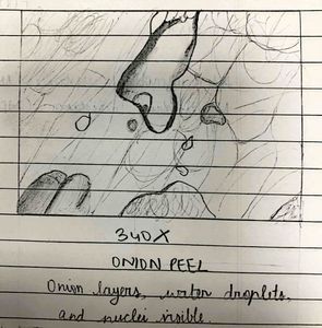
This is a picture and drawing of an onion peel under a 340X magnification.
We can see the cells of the onion are really rectangular and compactly packed. It looks somewhat like a brick wall. Upon closer inspection, we can see various irregularities present within such as varying cell sizes and shapes, development of bacteria etc.
The image with the dark irregular figure is the depiction of a bacterium in the onion. Around the cells and within it, we can see various particles such as nuclei, vacuoles, nutrients etc.
_____________________________________________ _______________________
Overall, the class today really showcased a different aspect of biology, which is very different from what I am used to studying. Looking into the various constituents of a cell through hands-on experience has definitely improved my experience and perspective about the subject.
Sign in to commentNobody has commented yet... Share your thoughts with the author and start the discussion!

 0 Applause
 0 Comments