The best of both worlds, where tradition and innovation meet.
 Apr 15, 2024 • 10:12 AM UTC
 South Africa
 140x Magnification
 Information
August H
Currently a postgraduate student at the University of Cape Town, focusing on differential gene expression in the developing brain.
2posts
0comments
1locations

View in Media Gallery
Kelps are a diverse group of multicellular aquatic organisms that fall under the classification of brown algae, making up the order Laminariales [1]. They are one of the most recognisable undersea entities, only eclipsed by the immense diversity of corals located in ecosystems such as the Great Barrier and Rainbow Reefs. As someone who grew up around the ocean, seeing these towering underwater behemoths anchored to the ocean floor, and the dried-up relics of these giants washed up along beaches was a common – and in many cases, fetid – occurrence. These seaweeds are easily mistaken as aquatic plants given their shared traits of “stems” and “leaves” however, they have distinct and specialised morphologies as a result of convergent evolution [2]. There are four main structural features typical of giant kelp: the holdfast that anchors the alga to the marine substrate, the stipe which resembles a stem (but does not transport nutrients, having a mainly supportive role), and gas bladders or pneumatocysts that contain air which allows for the blades of the kelp to remain close to the ocean surface to perform photosynthesis [3]. Being so accessible, they are an optimal target for both marine biologists and regular citizen scientists to study and elucidate their unique biology, of how their morphology and physiology are a result of the environment they exist in – and further, how the ecosystems they create benefit both aquatic biodiversity and coastal human communities.
Moving away from the exposition and onto the topic at hand, the Foldscope is a marvellous piece of equipment that brings the advancements of microscopy out of the sterile environments of research labs, and into the hands of curious-minded individuals who want to understand the world around them. An avenue of its use can be for researchers who aim to study samples collected from the field using microscopy, however, the process of gathering and preparing samples for imaging, and then only viewing them once provided access to a light microscope wastes time and prevents on-site analysis - this is where the Foldscope shines, allowing for this in media res data collection.
To illustrate the principle of collecting microscope data both on-site and in-lab, two sets of micrographs are presented. First, the following images are taken using the Foldscope of prepared kelp tissue sections.
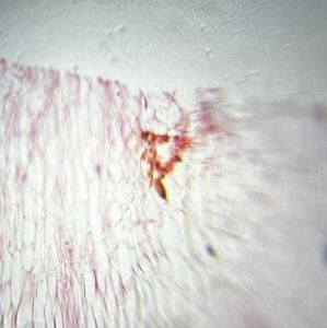
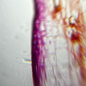
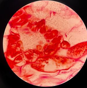
Despite their lower clarity, we can see the various cells and structures that make up the tissue section with enough detail to understand the microscopic organisation of the sample. This information can then be used as a stepping stone to decide what to focus on when analysis is performed in the lab. Homing in on the imaged structures, the following images are taken at 100x objective magnification.

In practice, the samples analysed using the Foldscope would not be prepared and stained and instead would look at larger structures or microscopic organisms, which is what the Foldscope is designed for. However, using an easy-to-operate, durable, and most importantly, portable microscope makes the data collection process more streamlined as the focus of the researcher can be on finding the best samples for collection by identifying them right on the spot. Additionally, from a student's perspective, going out into the field and collecting data becomes much more interactive, which overall enhances the learning experience.
In conclusion, being able to utilise the invaluable tools of microscopy both in the lab and out on research expeditions greatly enhances the ability of a researcher to gather vital data that neither one alone can provide. Therefore, the best use of the Foldscope, in my opinion, is in tandem with the traditional light microscope – the best of both worlds.
1. Starko, S., et al., A comprehensive kelp phylogeny sheds light on the evolution of an ecosystem. Molecular Phylogenetics and Evolution, 2019. 136 : p. 138-150.
2. S. T. Drobnitch, K.H.J., P. Prentice, and J. Pittermann, Convergent evolution of vascular optimization in kelp (Laminariales). Proceedings of the Royal Society B: Biological Sciences, 2015. 282 (1816).
3. Fowler-Walker, M.J., T. Wernberg, and S.D. Connell, Differences in kelp morphology between wave sheltered and exposed localities: morphologically plastic or fixed traits? Marine Biology, 2006. 148 (4): p. 755-767.
Sign in to commentNobody has commented yet... Share your thoughts with the author and start the discussion!

 0 Applause
 0 Comments