Microbial conflict in action: The fascinating world of amoebae
 Feb 17, 2023 • 11:23 PM UTC
 United States
 140x Magnification
 Microorganisms
Laks Iyer
Human observer of life. https://sukshmadarshin.wordpress.com
97posts
1255comments
5locations
View in Media Gallery
I've always been fascinated by amoebae. A single cell that does the work of a multicellular organism. The amoebal state is quite deceptive as across the tree of eukaryotic life, one sees it in many different branches, suggesting that it was an ancestral state.
Growing amoebae
Growing amoebae is fairly easy if you know what to feed it. I have maintained amoebae for months in a mixed culture containing a wheat grain as food. Typically, the wheat allows bacteria to go, which then feed some other flagellates which are consumed by amoebae. You essentially create a food chain. For this study though, I procured a commercial sample from Carolina Biologicals about 2 months back, Amoeba proteus and its prey Chilomonas . Chilomonas grows very easily when you drop a wheat grain into 20 ml of Springwater, whereas Amoeba proteus just needs Chilomonas to grow. It is easy to set up and needs to be reinoculated into fresh glassware (sometimes little glass bowls), every 2 weeks or so.
The goal
My goal was to make as many interesting observations of this amoeba. A lot of such observations are just waiting and watching. You set your foldscope up with the sample and watch for hours, perhaps a couple of days, usually between breaks for me. Today's smartphones allow one to make timelapse videos and can store a large volume of pictures and videos. A good number of them are just so but then suddenly you get a gem, often an unexpected gem and those are what I shall describe today. You should see this post by this high school girl. Wonderful videos of Amoeba proteus phagocytosing Chilomonas . https://microcosmos.foldscope.com/?p=transfer_174483
Inspired by this, I wanted to make some observations of my own and so I made a little chamber ( Click here on how to set up ) and put in a few amoebae and Chilomonas . These last for at least a couple of days.
The players in this ecosystem
Upon observing the sample, I realized that the culture wasn't a pure one and had a few other players. In reality, most of these commercial ciliate cultures have other contaminants that often add extra spice to the sample. Let us look at the four major players that were easily foldscopable in this slide.
1. Amoeba proteus , Chilomonas and a flagellate
View in Media Gallery
Also on youtube:
https://youtube.com/shorts/Ib3URrcqvAw?feature=share
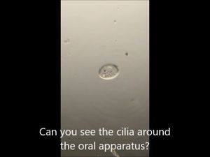
2. Vorticella . The anatomy of the Vorticella is quite nice in this video
https://youtube.com/shorts/Ib3URrcqvAw?feature=share
2. Vorticella . The anatomy of the Vorticella is quite nice in this video
View in Media Gallery
Also on Youtube
https://youtube.com/shorts/QoWLwKxPbhQ?feature=share
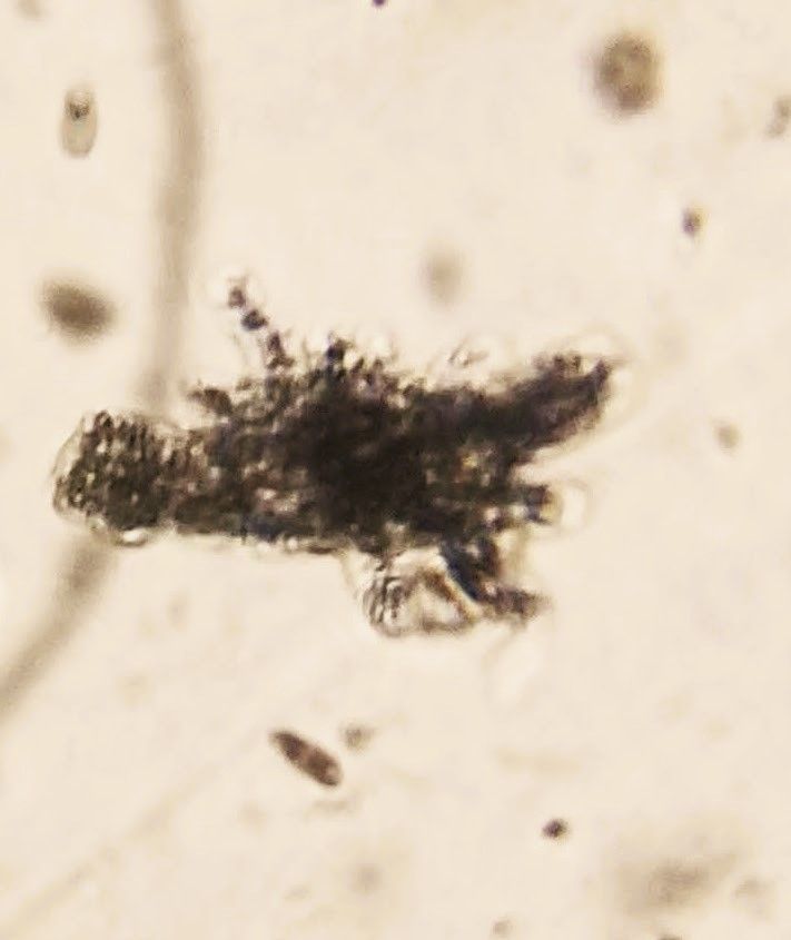
3. A little amoeba . My hypothesis is that these are amoeboflagellates. I suspect the little flagellate in the sample transforms into this amoeba. I didn't find the transforming step, but in my past observations, I have noticed several such amoeboflagellates.
https://youtube.com/shorts/QoWLwKxPbhQ?feature=share
3. A little amoeba . My hypothesis is that these are amoeboflagellates. I suspect the little flagellate in the sample transforms into this amoeba. I didn't find the transforming step, but in my past observations, I have noticed several such amoeboflagellates.
View in Media Gallery
https://youtube.com/shorts/33KMom7Ioxg
Beyond this of course a ton of bacteria that I haven't shown but you can see them at times in my videos of the sample.
Explorations
1. I was expecting to observe several phagocytosis events where Chilomonas are captured by the Amoeba , but to my surprise I caught this Amoeba chasing the little heterolobosean. These are time-lapse videos using the android app LapseIt pro. A picture is taken every 5 seconds in this case.
View in Media Gallery
https://youtube.com/shorts/a_PpwZz9HgA?feature=share
Thoughts
I find this quite remarkable and several points come to mind.
-Chemotaxis is definitely in play. What might the attractant/s be, some kind of small molecule like cyclic AMP?
- What might be the signaling pathways that reorient the larger amoeba to chase the smaller prey?
- If the little amoeba is truly a heterolobosean, then these are from very different branches of the tree of life, clearly, this is a pathway for lateral gene transfer in these unicellular organisms.
2. After some more hours of waiting, I finally caught this dramatic video where a voracious Amoeba phagocytoses a Chilomonas (right at the beginning), and then as though it needed some supplement it phagocytoses (plucks) the little amoeba.
View in Media Gallery
https://youtube.com/shorts/ouU_cdElP2M?feature=share
Thoughts
Again Chilomonas is from a completely different branch of life called SAR and I suspect the chemotactic molecule involved here is distinct from that one secreted by the little amoeba.
3. Finally here is a video where an amoeba repeatedly attempts to phagocytose a prey (it looks like a vorticella) with little effect. Either it is unable to wrest the sessile ciliate, or perhaps the prey/vorticella secretes something that is not palatable to the large amoeba.
View in Media Gallery
https://youtube.com/shorts/8jo8iSM43uw?feature=share
Thoughts
-Phagocytosis here involved repeated attempts to capture the same prey. How might a signaling network direct the amoeba to try again?
-If the prey has indeed found a way to reject the amoeba, one type of system that might be involved are conflict systems that the group I work for discovered and studies computationally (click here for references ). Might some of these be involved?
-The world of amoebae is fascinating, but there is a whole universe of chemistry involving small molecules, proteins, and perhaps other macromolecules in this that we don't see, and only guess for now. How might we unearth the answers to these deeper questions? We can ask several questions quite easily, but answering these would require some smart experimentation and of course funding. Perhaps the latter reason is why so many questions remain unanswered even after a hundred years of first observing these interesting phenomena in microbial life... Perhaps this is where frugal science could come into play and increase the space of the kind of experiments we can do from the comforts of our home?...
-Still observing this sample. Will post if something new comes up. To be continued.
Sign in to commentNobody has commented yet... Share your thoughts with the author and start the discussion!

 0 Applause
 0 Comments
_300x300.jpeg)