MtA_BIOL2201_2026 really cool pictures
 Apr 03, 2026 • 12:15 AM UTC
 Canada
 140x Magnification
 Plants
Jude Tapper
Learn about the author...
1posts
0comments
0locations
View in Media Gallery
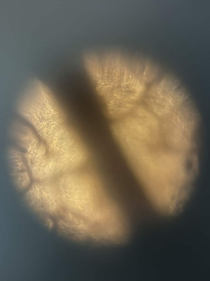
Below is an image of a dried leaf that displays a brownish-beige coloration, characteristic of tissue that has undergone seasonal changes and water loss. The vein-like structures extending outward from the stem are prominently visible, forming a network that is responsible for vascular tissue. In the spaces between these veins, a dotted pattern can be observed, which likely represents the remains of individual cells. The overall surface appearance is rough, brittle, and uneven, which makes sense with the breakdown of cellular integrity and the loss of moisture over time.

View in Media Gallery
This image below shows hair shed from my border collie mix, Wren, observed at 140x magnification using normal phone zoom. Because the hair is white, individual strands and follicles are clearly visible across the entire image. One noticeable follicle appears near the upper middle-left region, standing out due to its clarity and position. Along the length of the hairs, tiny spike-like projections can be seen, likely representing the cuticle scales that form the outer layer of each strand. The overall pattern consists of overlapping white strands, creating a layered, fibrous texture that fills the image..

View in Media Gallery
Sign in to commentNobody has commented yet... Share your thoughts with the author and start the discussion!
More Posts from Jude Tapper
No more posts from this author.