Day 31: A colonial vorticella: First explorations with fixed focus and dark field
 Nov 15, 2015 • 4:04 PM UTC
 Unknown Location
 140x Magnification
 Microorganisms
Laks Iyer
Human observer of life. https://sukshmadarshin.wordpress.com
97posts
1255comments
5locations
View in Media Gallery
Day 31: 11. Vorticella ( Alveolate->Ciliophora->Oligohymenophorea->Peritrichia->->->Vorticella)
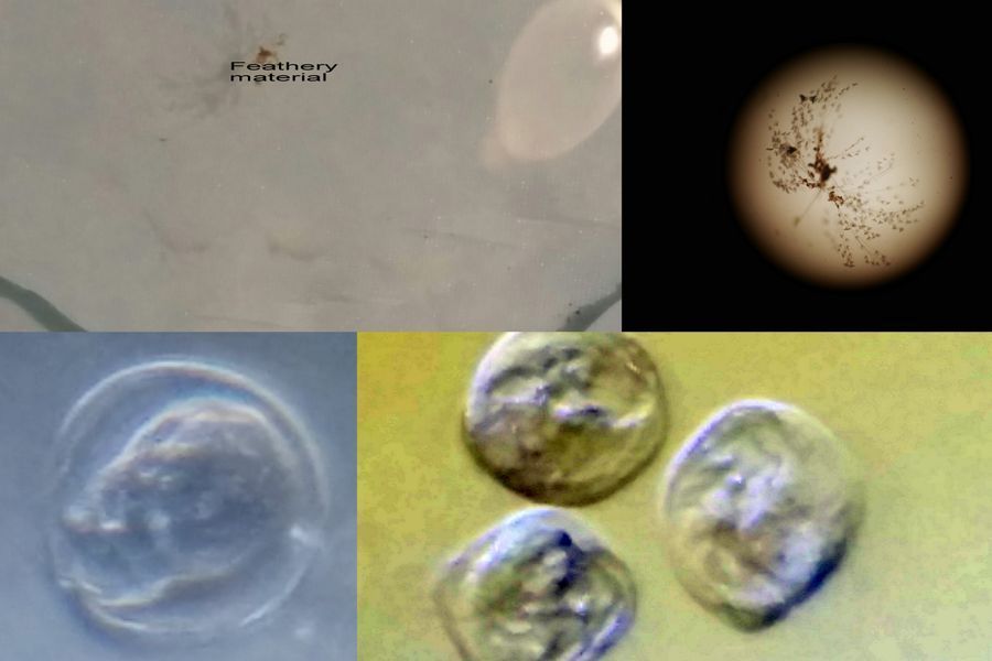
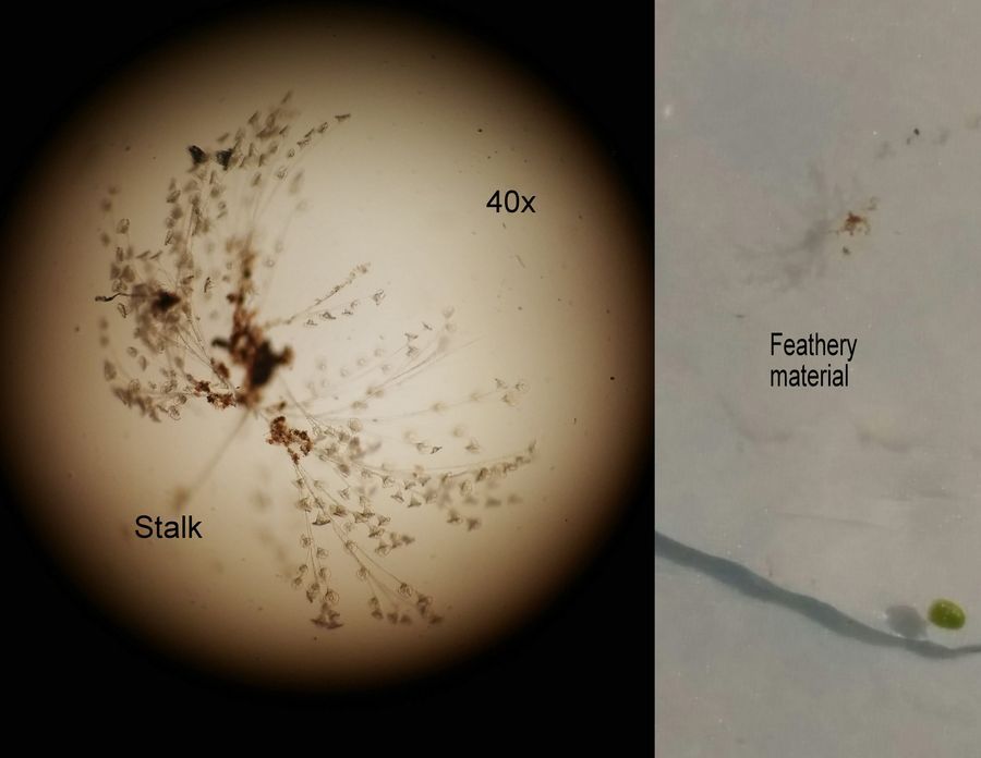
Ever since the first days of collecting the pond water sample ( Day 1 ), I have been observing this feathery object floating around. I always thought it was a feather fragment until today when I saw the object moving upwards and then in other directions almost with intention. I put it on a watch glass and looked at it under a low power microscope (40x). It was a pretty striking Vorticella colony. Here is a view at low power (40x) view. I wonder if you have read this wonderful post by Manu on the speed of the Vorticella spasmonemes .
Ever since the first days of collecting the pond water sample ( Day 1 ), I have been observing this feathery object floating around. I always thought it was a feather fragment until today when I saw the object moving upwards and then in other directions almost with intention. I put it on a watch glass and looked at it under a low power microscope (40x). It was a pretty striking Vorticella colony. Here is a view at low power (40x) view. I wonder if you have read this wonderful post by Manu on the speed of the Vorticella spasmonemes .
View in Media Gallery
Left. Vorticella at 40x. Right. The same on a watchglass. As you can see the Vorticellae are all connected like leaves to branches in a tree. The stalk is like the tree trunk and each Vorticella beats its cilia and the net movement is the result of the propeller-like action of all these vorticellae–Like a flying object with many propellers. Wonder if there is a signaling system between members of this colony (there has got to be one), that decides where to move. Normally we are taught that Vorticella have a motile phase where individuals move around and a sessile phase where they cluster together are are fixed. But here is an example of sessile Vorticella that move around as a group. I even took a movie of the same at low power. Manu, we need a 40x lens on the foldscope some day 🙂
However the most exciting thing was to use the newly rigged foldscope with dark field and fixed focus. This takes the cake (Thank you so much Foldscope team).
First foldscoping without all the new additions with my normal setup. This time I put a tape over the LED light source causing the light source to be more uniform. I really like the view.
First foldscoping without all the new additions with my normal setup. This time I put a tape over the LED light source causing the light source to be more uniform. I really like the view.
Now Foldscoping with fixed focus and dark field. The dark field needs some adjusting, but be assured in due course this will mature. You can see the beating patterns of the cilia more clearly in this. I filled up my phone with videos, so here are some excerpts (apologies for length, but I couldn’t contain myself). Fixed focus allows you to take movies or snapshots for as long as you want.
As you can see the Vorticella colony had broken up being squeezed by a water bubble in my slide(not shown) into the individuals (telotrochs?). I am still trying to figure out the optimal way of making slides for the foldscope with liquid samples. The most optimal one is that which can take a sizeable volume and not leak from the sides of the coverslip. Am going to try playing with coverslips separated by broken coverslips glass pieces. Also, a coverslip that can cover the whole slide would be more entertaining— working on all these.
Previous posts with this pond water sample: Day 1 , Day 6 , Day 17 , Day 27
Previous posts with this pond water sample: Day 1 , Day 6 , Day 17 , Day 27
Sign in to commentNobody has commented yet... Share your thoughts with the author and start the discussion!

 0 Applause
 0 Comments
_300x300.jpeg)


