The stratum corneum
 Nov 30, 2015 • 2:09 PM UTC
 Unknown Location
 140x Magnification
 Plants
Laks Iyer
Human observer of life. https://sukshmadarshin.wordpress.com
97posts
1255comments
5locations

View in Media Gallery
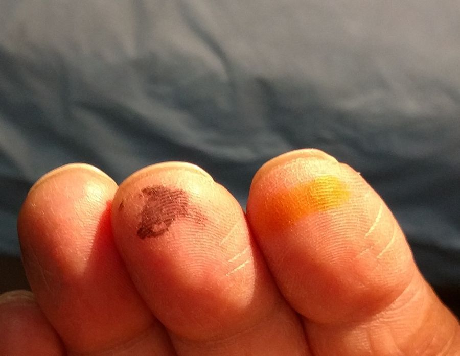
I have been wanting to do this experiment ever since I saw Manu’s post on volcanoes on his skin and the tatoo post . I essentially wanted to stain my skin and foldscope it. I had a somewhat erroneous idea that I might see some nucleated cells. I just had to do the experiment to see what would happen. It didnt start off as a planned experiment though. In fact, I was washing all my slides (100s or them) with soap, detergent and isopropanol. At the end of this uninspiring task, I thought staining my fingers would be a good way to round up the work. I used a desk lamp for illumination and observed them with a foldscope.
View in Media Gallery
The first thing that struck me was my peeling dry skin (Winter!). The pictures are from two different locations of my right hand, one from the finger tip and the other around a hair follicle. In both the peeling outer layer is obvious.

View in Media Gallery
In all three instances of finger staining, the stains fell into the grooves of the skin. Iodine formed little brown particles, the particles of India ink accumulated in the valleys of the grooves on my skin. Methylene blue that stains the nucleus showed interesting spreading patterns, possibly because it was dissolved in ethanol.
View in Media Gallery
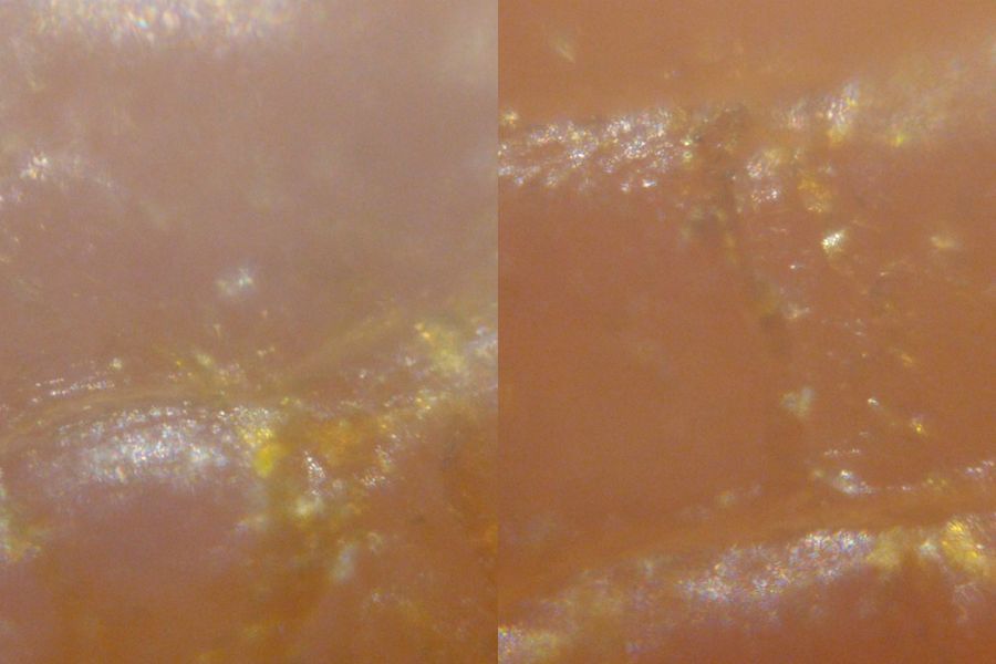
Finger stained with iodine
View in Media Gallery
Finger stained with India ink
View in Media Gallery
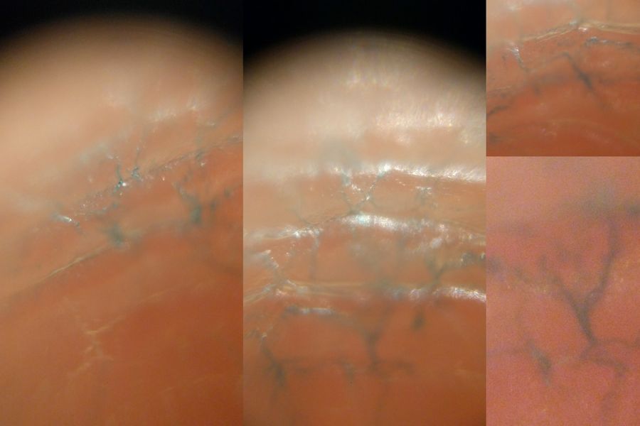
Finger stained with methylene blue These patterns illustrate the nature of the outher layer of the skin epidermis, called the stratum corneum. This is composed of encucleated dead cells which is about 15-20 cells thick. Starting from the stratum basale, they migrate up to the top in 14 days to form the stratum corneum. The cells are highly keratinized, without a nucleus or other organelles. Perhaps a protein stain might have been interesting. The patterns also give an idea of the skin surface, one of our greatest innate defensive systems.
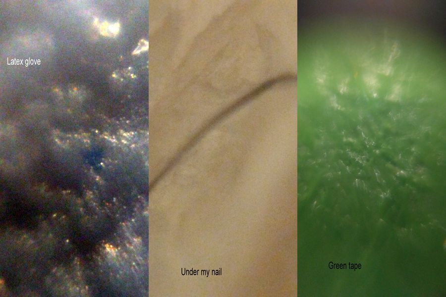
Foldscopes have tremendous scope for surface views. Here are a few more surface views. Pictures are labeled.
Foldscopes have tremendous scope for surface views. Here are a few more surface views. Pictures are labeled.
View in Media Gallery
Surface views of Latex gloves, underneat my fingernail (ugh!) and green tape.
Sign in to commentNobody has commented yet... Share your thoughts with the author and start the discussion!

 0 Applause
 0 Comments
_300x300.jpeg)


