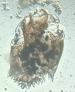
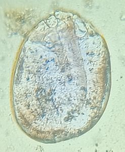
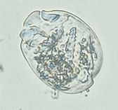
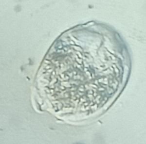
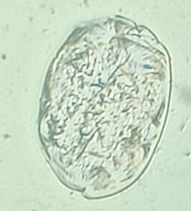
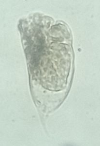
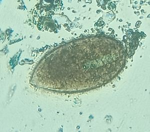
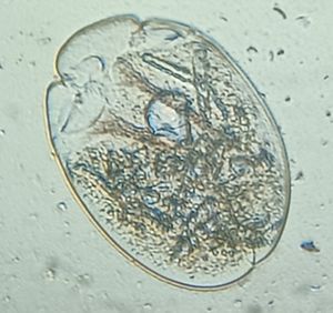
Amphistome egg
 Feb 06, 2019 • 3:57 AM UTC
 Unknown Location
 140x Magnification
 Microorganisms
Meignanalakshmi Sundaram
I am Dr.S.Meignanalakshmi, working as Professor, at the Directorate of Centre for Animal Health Studies, TANUVAS, Chennai-51. Working on Foldscope project on "Foldscope for diagnosis of Rumen Acidosis and parasitic infections in cattle" sanctioned by DBT
66posts
8comments
1locations

View in Media Gallery
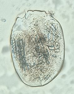
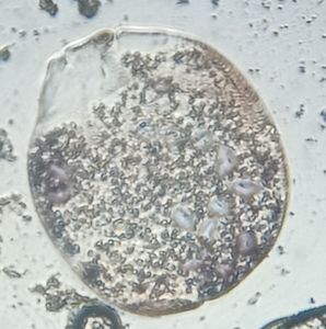
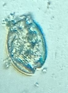
2-3 g of dung sample was taken, mixed with water and then sieved. The mixture was centrifuged at 2000 rpm for about 2 minutes. The supernatant was discarded and a drop of the sediment was placed on a slide with a cover clip on top. It was then examined under foldscope. It was observed that the Amphistome egg is colorless, oval in shape with composite yolk (figure 1)

View in Media Gallery
Amphistome egg
Sign in to commentNobody has commented yet... Share your thoughts with the author and start the discussion!

 0 Applause
 0 Comments