Experiments with blood-II. Staining white Blood Cells
 Jan 18, 2015 • 11:25 PM UTC
 Unknown Location
 140x Magnification
 Microorganisms
Laks Iyer
Human observer of life. https://sukshmadarshin.wordpress.com
97posts
1255comments
5locations
View in Media Gallery
A common refrain I read in articles by amateurs is that every microscopist should at least have eosin Y and methylene blue on their shelves. Methylene Blue is commonly sold by aquarium shops and eosin Y can easily be obtained at a small price (I bought it on the internet). My previous attempt to stain blood smears with alcohol fixing and methylene blue gave satisfactory results for RBCs but WBCs were very disappointing. See https://microcosmos.foldscope.com/2015/01/14/experiments-with-blood/ . The standard blood stains are a mixture of a variant of methylene blue and Eosin Y, which are combined either in a buffered soluton or methanol. I thought I’d just mix the two in equal proportion and add them to the smear and see what happens.
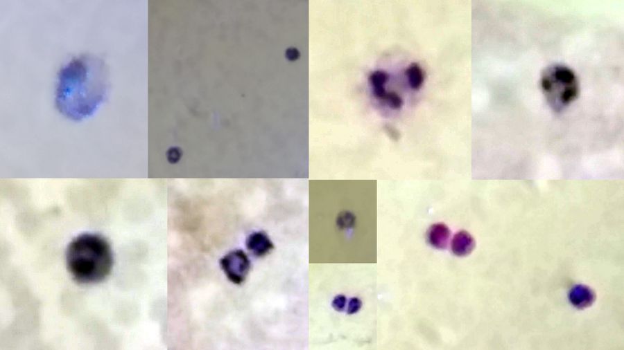
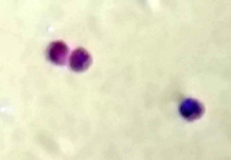
So for this experiment, I fixed my cells with Rubbing alcohol (70% ethanol) for a minute and stained with a 1 % Eosin in aqueous solution and Methylene Blue mixture for 5 minutes and washed it off with tap water. This time I used the foldscope tape instead of the coverslip over the smear. The RBCs were not very visible, but I did find many WBCs to my delight. These are just slightly larger than the RBCs (faint background). I could also find some of the major types of WBCs. These are distinguished by the structure of their nucleus and color of the cytoplasm.
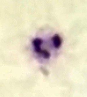
1. The Neutrophil is distinguished by a nucleus with multiple DNA lobes (typically 4), which comprises over 50% or more of the WBCs.
So for this experiment, I fixed my cells with Rubbing alcohol (70% ethanol) for a minute and stained with a 1 % Eosin in aqueous solution and Methylene Blue mixture for 5 minutes and washed it off with tap water. This time I used the foldscope tape instead of the coverslip over the smear. The RBCs were not very visible, but I did find many WBCs to my delight. These are just slightly larger than the RBCs (faint background). I could also find some of the major types of WBCs. These are distinguished by the structure of their nucleus and color of the cytoplasm.
1. The Neutrophil is distinguished by a nucleus with multiple DNA lobes (typically 4), which comprises over 50% or more of the WBCs.
View in Media Gallery
2. The Basophil is distinguished by a strongly blue cytoplasm with a bilobed nucleus.
View in Media Gallery
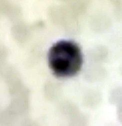
3. The Monocyte is distinguished by a Kidney-shaped nucleus.
View in Media Gallery
I did find lymphocytes too but didnt get a nice picture. Might post one later.
Future directions:
1. I need to get a better fixing solvent so that cells (esp. RBCs) dont get mis-shaped. It is interesting that one can be very careless with cheek cells, and get very good staining without fixing. I have been able to keep the cheek cell slides for over a week now. In contrast RBCs are very delicate and very sensitive to the solutions they are exposed to. If the fixing is not too good, the cells quickly fuse, become mis-shaped and form clumps. Methanol is a standard fixing solvent used by experts. Since I havent found an off-the-shelf version, I have been using denatured alcohol or rubbing alcohol. Need to try heat fixing next time. Clearly when it comes to staining cells even from the same organism, there are different strokes for different folks.
2. There are alternatives to Eosin and Methylene blue and people have tried food dyes, henna, tumeric extracts. One of my favorite microscopists, the late Walter Dioni gives many ideas for alternative stains. http://www.microscopy-uk.org.uk/mag/indexmag.html?http://www.microscopy-uk.org.uk/mag/artmay11/wd-Brilliant-Blue-1.html. I wonder if any of these can give good contrasts like the standard blood stains.
Foldscope notes: I was keen to explore this in the high-mag, but ever since the lens holder gave way, all my attempts to get a nice image have proved futile. Hopefully, I can get it to work satisfactorily soon. Manu, I cannot see the image clearly when I load the lens above the stage, but it seems to work when fixed below, but it is harder to take pictures with as centering seems to be an issue. There is also a polarity in which side faces the sample and which side the eye (Should it really matter for these?). As with all these, trials and errors are likely to solve these issues.
In all this with the bad hands I have, I also tore the holder of the 140x lens while peeling it off the stage. I have managed to fix it with glue, with my mutilated high-mag lens holder as a holder.
Sign in to commentNobody has commented yet... Share your thoughts with the author and start the discussion!

 0 Applause
 0 Comments
_300x300.jpeg)


