Watching inside the brain of a live ant!! Part 1
 Nov 20, 2014 • 4:30 AM UTC
 Unknown Location
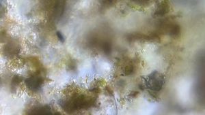
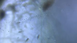
 140x Magnification
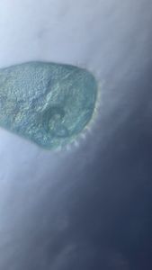
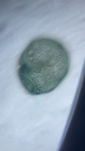
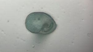
 Microorganisms
Manu Prakash
I am a faculty at Stanford and run the Prakash Lab at Department of Bioengineering at Stanford University. Foldscope community is at the heart of our Frugal Science movement - and I can not tell you how proud I am of this community and grassroots movement. Find our work here: http://prakashlab.stanford.edu
266posts
1198comments
42locations
View in Media Gallery
Summary: I have been fascinated with the idea of watching live brains. They are the most complex machines that we know (maybe in the universe). Here is a protocol to get you started to image live “ant brains”
Ants are everywhere. It does not matter if you like them or you don’t. They steal your mini-sugar cubes, inspire you with diligence and hard work and are prime examples of cooperative behavior (almost like a super organism of some kind). So anywhere you are, you probably have an ant crawling around you somewhere nearby. So why not take a look at a live ant; sounds like a project.
I wanted to make sure the ant is alive, but I wanted it to be still enough so I can watch it carefully under a Foldscope. Again, we use our common trick of trapping an ant between two transparent tapes without squashing it. It’s just sticky enough between the tapes, that the ant will stay immobilized and we can watch the innards.
Methods:
1. Catch an ant that you like. You will soon realize, so many different species of ants exist on this planet. They have all kinds of morphologies that are distinct. So choose your favorite one.
2. It might be ideal to take a few pictures of the ant before you get started. This will help you ID the species of ant you are working with.
3. Don’t pick the ant with your hand. Most likely you can damage it; although they are pretty hardy to begin with. I usually use tape and just being the tape close to the ant. By gently touching the ant, it gets stuck on the tape. Now quickly, I take the second piece of clear tape and use this method to mount the ant in our a foldscope slide.
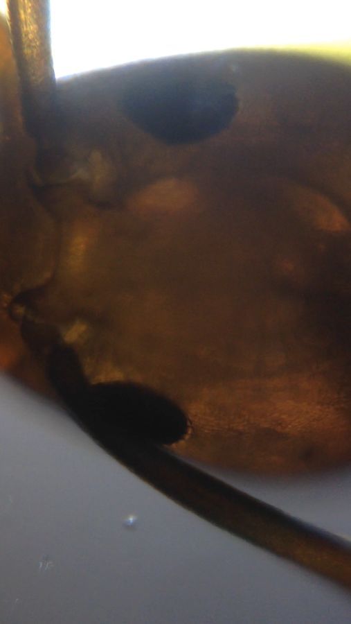
4. I decided to image the ant with my 140X Foldscope coupled to an iphone5 camera in video mode.
http://youtu.be/ho6AGSD_Ck8
Results and observations:
1. You can see the first videos I took. Also notice the energy and excitement in my voice – because I was not expecting that the ant was could be so transparent and I could actually see inside. That was quiet a thrill. See video below.
2. You can easily notice the tracheal structures that supply oxygen to the head (and the brain). You can notice a loop of tracheal strictures branching to the right and the left, and joining together.
3. What is most fascinating and apparent immediately (I have done this twice and found it to be true in both samples); is a kind of a pulsatile movement inside the head. This is exciting and can only be seen in live imaging (watch video carefully).
4. Two possibilities exist for the live pulsations/oscillations in the head – one could be fluid pump or pressure pulses from the hemolymph (ants and all insects have many hearts, and it could generate a pressure – just like our own pulses/beats). Second option could be that what we are observing is a jaw muscle movement.
5. While imaging the ant, I also took videos of the jaws of an ant. I will post them some other time.
Next steps:
1. This is only the tip of an iceberg – having optical access to the brain is Very exciting. I will definitely focus on this part first and look at some markets to identify the brain tissue (by marking some of the lipids).
2. The pulsations are very intriguing and I need to figure out where they really come from.
3. A simple stain for muscles might also do the trick – I will try by injecting some stains for the same as well.
4. I need to do this for more number of species; let’s see what type of signals I get for other species.
Ants are everywhere. It does not matter if you like them or you don’t. They steal your mini-sugar cubes, inspire you with diligence and hard work and are prime examples of cooperative behavior (almost like a super organism of some kind). So anywhere you are, you probably have an ant crawling around you somewhere nearby. So why not take a look at a live ant; sounds like a project.
I wanted to make sure the ant is alive, but I wanted it to be still enough so I can watch it carefully under a Foldscope. Again, we use our common trick of trapping an ant between two transparent tapes without squashing it. It’s just sticky enough between the tapes, that the ant will stay immobilized and we can watch the innards.
Methods:
1. Catch an ant that you like. You will soon realize, so many different species of ants exist on this planet. They have all kinds of morphologies that are distinct. So choose your favorite one.
2. It might be ideal to take a few pictures of the ant before you get started. This will help you ID the species of ant you are working with.
3. Don’t pick the ant with your hand. Most likely you can damage it; although they are pretty hardy to begin with. I usually use tape and just being the tape close to the ant. By gently touching the ant, it gets stuck on the tape. Now quickly, I take the second piece of clear tape and use this method to mount the ant in our a foldscope slide.
4. I decided to image the ant with my 140X Foldscope coupled to an iphone5 camera in video mode.
http://youtu.be/ho6AGSD_Ck8
Results and observations:
1. You can see the first videos I took. Also notice the energy and excitement in my voice – because I was not expecting that the ant was could be so transparent and I could actually see inside. That was quiet a thrill. See video below.
2. You can easily notice the tracheal structures that supply oxygen to the head (and the brain). You can notice a loop of tracheal strictures branching to the right and the left, and joining together.
3. What is most fascinating and apparent immediately (I have done this twice and found it to be true in both samples); is a kind of a pulsatile movement inside the head. This is exciting and can only be seen in live imaging (watch video carefully).
4. Two possibilities exist for the live pulsations/oscillations in the head – one could be fluid pump or pressure pulses from the hemolymph (ants and all insects have many hearts, and it could generate a pressure – just like our own pulses/beats). Second option could be that what we are observing is a jaw muscle movement.
5. While imaging the ant, I also took videos of the jaws of an ant. I will post them some other time.
Next steps:
1. This is only the tip of an iceberg – having optical access to the brain is Very exciting. I will definitely focus on this part first and look at some markets to identify the brain tissue (by marking some of the lipids).
2. The pulsations are very intriguing and I need to figure out where they really come from.
3. A simple stain for muscles might also do the trick – I will try by injecting some stains for the same as well.
4. I need to do this for more number of species; let’s see what type of signals I get for other species.
Sign in to commentNobody has commented yet... Share your thoughts with the author and start the discussion!

 0 Applause
 0 Comments