Red Onion Cells
 Dec 31, 2015 • 6:27 AM UTC
 Unknown Location
 140x Magnification
 Unknown
P. Mooney
Learn about the author...
6posts
2comments
1locations
View in Media Gallery
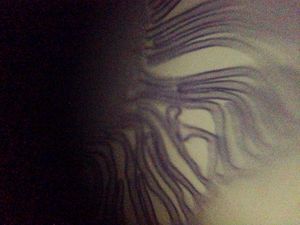
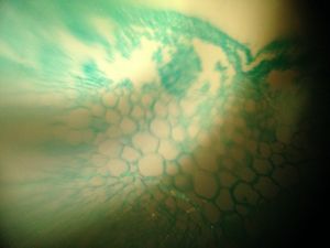
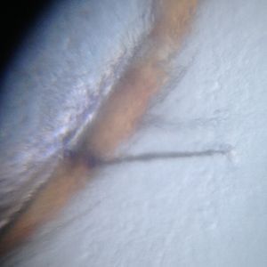
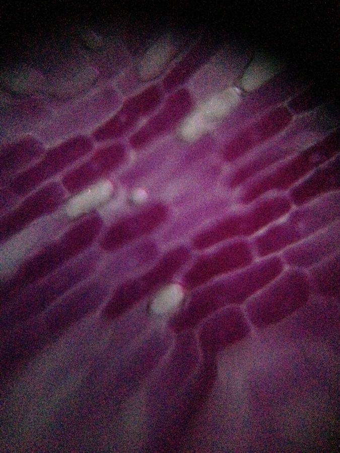
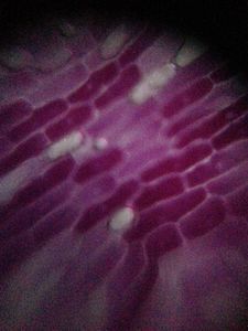
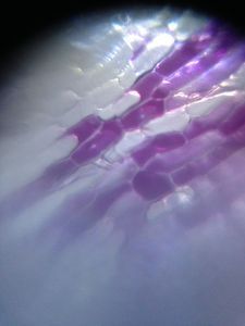
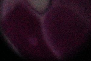
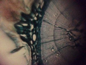
A thin section of a red onion (one layer beneath the outermost layer) was sandwiched between two layers of transparent packing tape. Next, the onion was imaged with a FoldScope that was equipped with a low-magnification (140x) lens, LED light module, and iPhone (5) camera. In this manner, it was possible to observe individual onion cells (see attached images). Interestingly, many of these onion cells contained small circular structures that were located near the center of the cell. I suspect that they may be nuclei. An additional image of a candidate nuclei was acquired using a high-magnification (500x) lens. In the future, it would be interesting to use a nuclei-specific stain in order to determine if the small circular structures are indeed nuclei. Furthermore, it would be interesting to see if image quality might be increased by using only a single layer of transparent packing tape.
Sign in to commentNobody has commented yet... Share your thoughts with the author and start the discussion!
 0 Applause
 0 Comments