Strange ciliate of Drumheller Fountain (Frosh Pond), University of Washington
 Jan 11, 2016 • 8:34 AM UTC
 Unknown Location
 140x Magnification
 Microorganisms
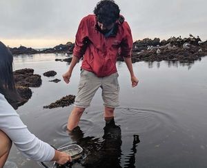
Manu Prakash
I am a faculty at Stanford and run the Prakash Lab at Department of Bioengineering at Stanford University. Foldscope community is at the heart of our Frugal Science movement - and I can not tell you how proud I am of this community and grassroots movement. Find our work here: http://prakashlab.stanford.edu
266posts
1198comments
42locations

View in Media Gallery
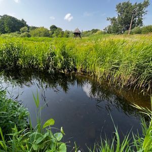
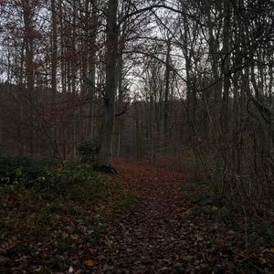
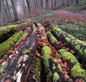
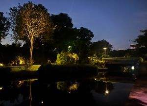
I was kindly invited to give a talk at University of Washington (EE department) campus last week. I had a wonderful (although hectic) 24 hours. In between my meetings and running from one office to another; I squeezed an early morning moment to stop at this traditional U.Washington sight; the Drumheller Fountain. It’s got some colorful history – but needless to say, the geese in this pond is quiet bold. This is visible in it;s attempt to feed on me while I tried to sample the waters. I sampled the water with my host of the day, Prof. Matt Reynolds – who will soon be a Foldscoper himself 🙂
So, what’s fun about sampling a large fountain that is almost a 100 years old is the microscopic life forms. I am not sure if this pond dries out anytime of the year (how can anything dry out in Seattle) – but I find it very interesting to think that single cell ciliates living in the pond might have survived generation after generation for more than 100 years. Clearly, I don;t know this – but all I can do; is dive in this wonderland and look for what I find.
I note that the water was quiet cold.
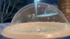
As usual, when I do higher resolution imaging, I always use glass slides and cover slips. This is the best way to get highest resolution in your imaging.
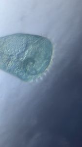
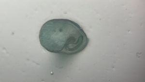
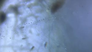
Here is the first wonderful surprise; a ciliate that I caught for almost 5 minutes. I can observe some really strange things which I should be able to use to identify the same; including an isometric body; with a curved tip. This ciliate has a capacity to “turn on a dime” this usual rotation is employs very often. I am still going through “Kahl’s ciliate book” to identify this beast; but that should not stop me from sharing this with everyone. Maybe someone else can identify this for me.
I note that the water was quiet cold.
As usual, when I do higher resolution imaging, I always use glass slides and cover slips. This is the best way to get highest resolution in your imaging.
Here is the first wonderful surprise; a ciliate that I caught for almost 5 minutes. I can observe some really strange things which I should be able to use to identify the same; including an isometric body; with a curved tip. This ciliate has a capacity to “turn on a dime” this usual rotation is employs very often. I am still going through “Kahl’s ciliate book” to identify this beast; but that should not stop me from sharing this with everyone. Maybe someone else can identify this for me.
This is the first time I have even been able to image the same swimming microorganism for as long as 5 minutes. It is clear to me that some kind of artificial trapping mechanism implemented in the glass slide will be wonderful for actually imaging ciliates for long duration of time. I have several attempts ongoing – including a clever way to coat an entire glass slide with a criss cross pattern of nail polish – and covering the top with cover slip. Results of this experiment will be reported soon. You could beat me to the race and share your own “ciliate jail” sooner..
A crucial point here is that I recorded the same organism for almost 5 minutes. This way I am able to see repeated behaviors in the same organism over long enough period of time to start making some inferences and observations. One that I am the most happiest about is the fact that since the ciliate has a flat bottom and curved top (thus dorsal (top) and ventral (bottom) surface). Since this is a ciliate, I am assuming it is covered in cilia. Now, when the bottom flat surface is in contact with the substrate – I believe the organism is only capable of making “clock wise” turns. I have looked carefully (since in the video you see both clockwise and anti-clockwise turns); but every time a clockwise turn happens; the flat surface contacts the top cover slip – while when an anti-clockwise turn happens, the ventral surface of the ciliate is touching the bottom surface. This would be a really fun fact; since it would mean that the ciliary covering is chiral (which is a common feature in many ciliates). I just also like the idea of driving around a car that could only turn right (old RC cars used to be like that); and if you want to move left; you just turn right.. a lot.
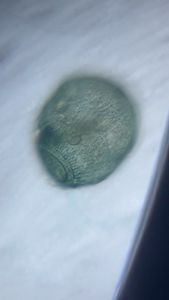
I also found several other characters which were also intriguing, including a similar looking ciliate – albeit much smaller.
A crucial point here is that I recorded the same organism for almost 5 minutes. This way I am able to see repeated behaviors in the same organism over long enough period of time to start making some inferences and observations. One that I am the most happiest about is the fact that since the ciliate has a flat bottom and curved top (thus dorsal (top) and ventral (bottom) surface). Since this is a ciliate, I am assuming it is covered in cilia. Now, when the bottom flat surface is in contact with the substrate – I believe the organism is only capable of making “clock wise” turns. I have looked carefully (since in the video you see both clockwise and anti-clockwise turns); but every time a clockwise turn happens; the flat surface contacts the top cover slip – while when an anti-clockwise turn happens, the ventral surface of the ciliate is touching the bottom surface. This would be a really fun fact; since it would mean that the ciliary covering is chiral (which is a common feature in many ciliates). I just also like the idea of driving around a car that could only turn right (old RC cars used to be like that); and if you want to move left; you just turn right.. a lot.
I also found several other characters which were also intriguing, including a similar looking ciliate – albeit much smaller.
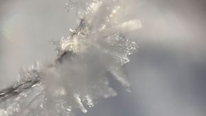
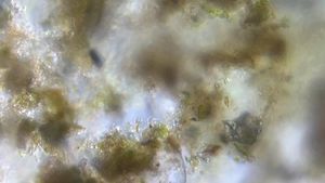
I was able to image this ciliate for a long time, since it self-trapped itself under a very fine decaying bacterial/fungal film. Or this could potentially be a leaf epidermis where only cell wall remains. The peculiar pattern tells me it might be a remaining plant cell wall of a leaf that de-generated. But I have been looking for fungal hype networks for some time – so that is another real possibility.
Finally, a nematode also showed up with a collection of several other ciliates.
And what pond would complete without a rotifer of it’s own. I was proud of the fact that I was able to image this one quiet clearly, and so I should be able to identify it. Specially because of the fact that I only have ~4000 possible candidates to go through.
Finally, I also encountered another ciliates very commonly. I am still in the process of identifying these guys still (please leave comments, make observations below). But I often found them in couples or triplets.
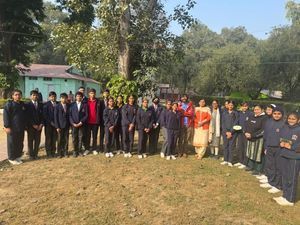
Now, at the end of my lecture – I also left some foldscope for UW students. So here is a challenge – what else can you find in your beloved pond? Also; can someone actually confirm that in the last 100 years of the history of this fountain, it has never dried out. Even if it has dried out, many spores will survive. What I would be more worried about is treating the water – I hope that in the future, they don’t clean this fountain ever again. For the sake of visitors at the least.
cheers
manu
ps: this post would not have been possible without the my generous host, Prof. Matt Reynolds. Thanks for inviting me to rainy Seattle. It was an incredible day, and I learned a lot.
cheers
manu
ps: this post would not have been possible without the my generous host, Prof. Matt Reynolds. Thanks for inviting me to rainy Seattle. It was an incredible day, and I learned a lot.
Sign in to commentNobody has commented yet... Share your thoughts with the author and start the discussion!
 0 Applause
 0 Comments