Moth Observation – Bio60_2019
 Feb 03, 2019 • 8:14 PM UTC
 Unknown Location
 140x Magnification
 Unknown
Claire Jordan
Learn about the author...
2posts
0comments
1locations
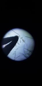
This image depicts a large, circular black mass, which I took to be the moth’s eye. The image quality is not ideal due to the large size of the section and zoom capacity of the foldscope. After noticing a pesky moth flying around my room, I decided to observe it with my foldscope. After catching the moth in an empty container, being careful not to crush it, I placed the insect in the freezer, effectively kill it and increasing the ease of the dissection process. I initially hoped to look at the moth’s wing under the microscope; however, I did not anticipate the difficulty of handling moth wings. Even after I cut one off, the section appeared too opaque for observation.
View in Media Gallery
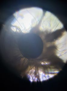
This image depicts the moth’s bent front leg, just below its eye. I placed the remaining section on a microscope slide and placed two pieces of tape on either side of the sample to ensure that I didn’t crush the moth when I positioned the cover slip. The resulting images appear slightly dark and blurry, which I attribute to the girth of the moth sample. With more precise tools, I would have liked to cut the moth into thin sections to have better visibility of individual cells.
View in Media Gallery
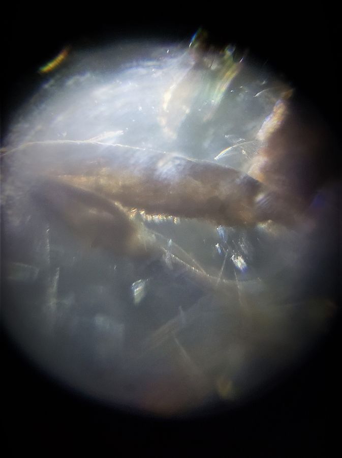
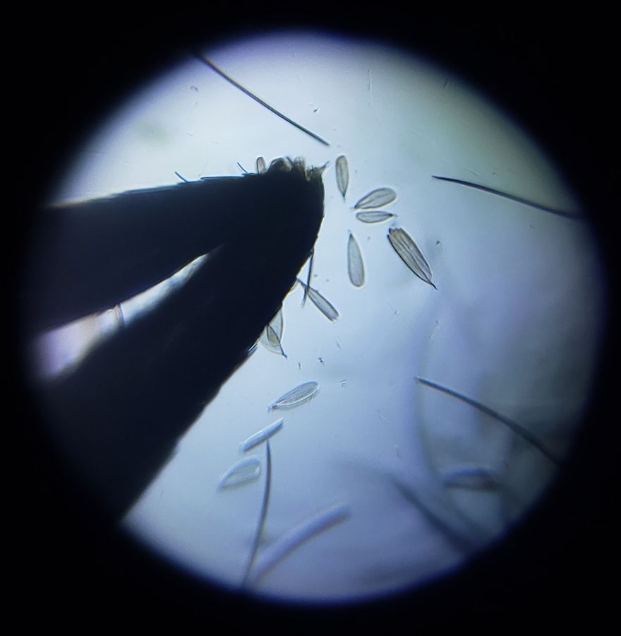
I was surprised to discover, when positioning the moth, how easily the moth’s legs stuck to the tweezer. The image depicts one of the moth’s joints, which I had been using to rotate the moth on the slide. The flake-like sections appear to be small pieces of the leg that I accidentally brushed off. Their translucence is a stark contrast to the dark, solid appearence of the leg.
Sign in to commentNobody has commented yet... Share your thoughts with the author and start the discussion!

 0 Applause
 0 Comments