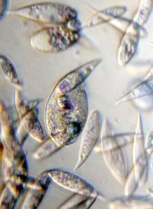
Tetrahymena thermophila mutants: Fat, Mouthless and Balloon
 Feb 11, 2019 • 11:23 PM UTC
 Unknown Location
 140x Magnification
 Microorganisms
Laks Iyer
Human observer of life. https://sukshmadarshin.wordpress.com
97posts
1255comments
5locations

Thanks to the wonderful ASSETT program , I had the opportunity to procure various temperature sensitive mutants of Tetrahymena thermophila . These grow normally at room temperature, but at 37℃ /98.6℉ they display defects due to mutations in genes involved in cell division (hence called temperature sensitive mutants).
My first step in reproducing these phenotypes was to find a way to make a 37℃ /98.6℉ incubator in my home lab. Following is a description of my low-tech hack. The culture tubes were placed in a water bath (a plastic bowl) which was further placed under an inverted glass jar. The glass jar was heated using a light bulb that was further controlled using a digital thermostat. I repurposed the thermostat from a heating mat that I use regularly to germinate seeds in winter (see heating mat) .
My first step in reproducing these phenotypes was to find a way to make a 37℃ /98.6℉ incubator in my home lab. Following is a description of my low-tech hack. The culture tubes were placed in a water bath (a plastic bowl) which was further placed under an inverted glass jar. The glass jar was heated using a light bulb that was further controlled using a digital thermostat. I repurposed the thermostat from a heating mat that I use regularly to germinate seeds in winter (see heating mat) .

The cultures were first grown for a couple of days in NEFF medium and then were incubated overnight at 37℃ /98.6℉ (temperature shift). I would love to hear of other incubator-design hacks from the community. One which is portable would make it even cooler.
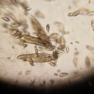
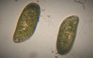
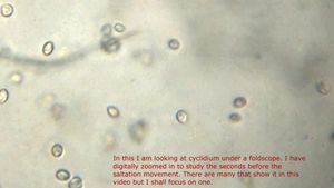
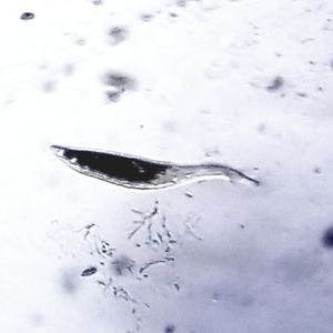
Following videos show the division mutants. First let us look at wild type Tetrahymena thermophila under a foldscope so as to get a comparative base.
Following videos show the division mutants. First let us look at wild type Tetrahymena thermophila under a foldscope so as to get a comparative base.
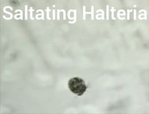
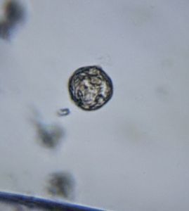
Now let us look at the mutant called Fat. Here the cell becomes round and “fat”, and shows an abnormal swimming pattern. Note that in this video, the cells have been slowed down with a detaining solution. Also video starts after 10s or so.
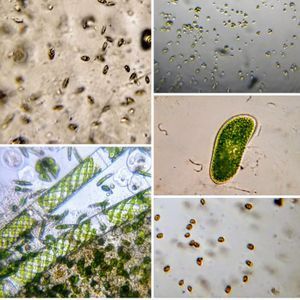
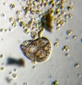
This mutant is called Mouthless as it doesnt have a mouth. In this instance the population was low, perhaps as I had it at the higher temperature so long that most cells just died. Mutant spherical cells are seen.
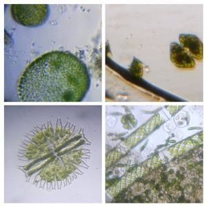
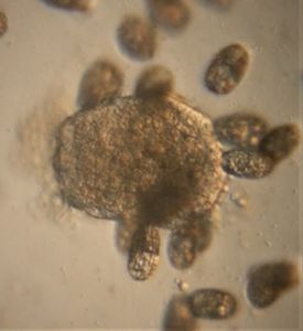
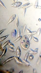
This is by far the most spectacular of the mutants and is called Balloon. I think the Balloon mutant is telling us something important — which is the range of ciliate shapes shouldnt come as a surprise. Here a single mutant shows a variety of shapes. Perhaps ciliate shape diversity could involve polymorphisms in such genes? A corollary to that would be that several ciliates might not be as different from each other as their shapes suggest.
Genetic mutants played a big role in understanding the biology of an organism, they still do. Thousands of mutants in model organisms changed the way we understood the biology of various life forms. However, in the past most mutants were isolated either naturally or by exposing cells to mutagens and selecting mutants. Now with genome sequences, we can alter specific genes and look for phenotypes. However, mutants still provide deep insights, so it is important to keep looking carefully and you might be on to something interesting if you find a mutant.
Sign in to commentNobody has commented yet... Share your thoughts with the author and start the discussion!
 0 Applause
 0 Comments
_300x300.jpeg)
