Is it Magic? De-mystifying the Foldscope
 Feb 18, 2019 • 1:13 AM UTC
 Unknown Location
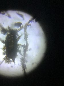
 140x Magnification
 Unknown
Jayashree Ramadas
We are a group of students, volunteers and staff working with TIFR Hyderabad's Science Education and Outreach program: http://www.tifrh.res.in/~outreach/
39posts
26comments
2locations
View in Media Gallery
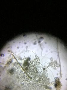
Recently in a workshop we probed into students’ curiosity related with their experiences with Foldscopes. One particular question, asked by a student of Grade 9, sparked a lively discussion in the workshop. The question was simple yet striking: “ Is there any magic in the Foldscope? “, the student asked. She was perhaps curious to know how such a simple, inexpensive, small paper-folded device is able to magnify as much as a heavy and expensive compound microscope. Another student, from Grade 8, was more specific: “ What is the lens used in the Foldscope and how can we make our own Foldscope? ” We realised there was a sense of mystery among the students regarding the mechanism of Foldscopes. Their understanding of simple and compound microscopes did not gel with their Foldscope experiences.
Starting with how Leeuwenhoeck observed the details of cells and even bacteria using a single lens and a well designed platform, we found ourselves harking back to concave and convex lenses, real and virtual images, the critical parameter of distance of object from the lens (comparing with a spoon as a mirror), diagrams to explain that, ‘ more the curvature of a convex lens, larger is the image ’, and the challenges of working with a spherical, or ball lens . Parts of the compound microscope were shown that are anologous to the Foldscope — the slide stage with slide slot and coarse to fine adjustment with the focus ramp.
Starting with how Leeuwenhoeck observed the details of cells and even bacteria using a single lens and a well designed platform, we found ourselves harking back to concave and convex lenses, real and virtual images, the critical parameter of distance of object from the lens (comparing with a spoon as a mirror), diagrams to explain that, ‘ more the curvature of a convex lens, larger is the image ’, and the challenges of working with a spherical, or ball lens . Parts of the compound microscope were shown that are anologous to the Foldscope — the slide stage with slide slot and coarse to fine adjustment with the focus ramp.

View in Media Gallery
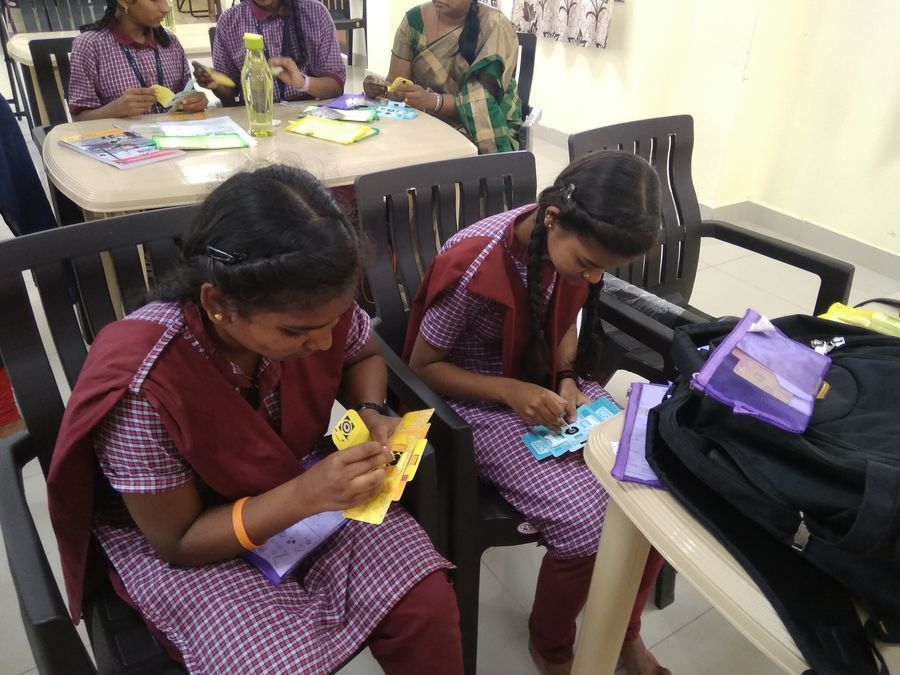
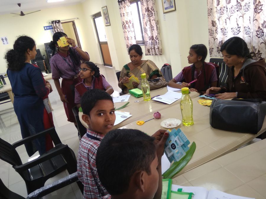
Discussing the mechanism of Foldscope, comparing with a compound microscope That’s when students realised that their Foldscope consists of only one ball lens with magnification of 130X. There was wonder on their faces after checking their Foldscopes, realising that it is an engineering innovation by the inventors (Manu and Jim) to design the accurate folding of the Foldscope to hold the slide, move it and then bring the lens close to the sample to obtain clear magnified images, comparable to a compound microscope.
View in Media Gallery
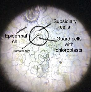
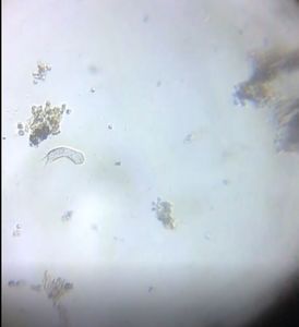
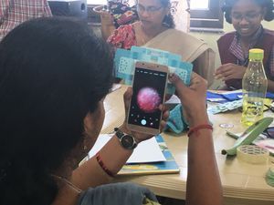
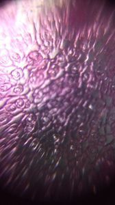
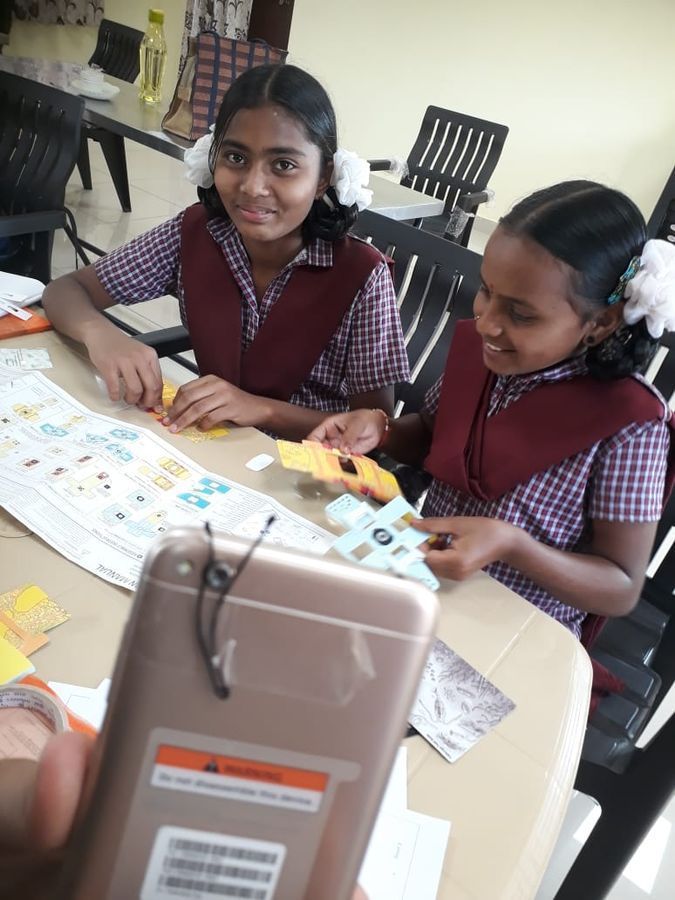
Students checking the lens in their Foldscopes While the Foldscope was being demystified, one of the teachers demonstrated an innovative technique, to attach the lens of a laser pointer to a cell phone camera with a hairclip, which she found on YouTube . We observed the epidermal layer of a leaf using this hairpin microscope and clearly saw the cells and the stomata!! It was an exciting moment for all of us and especially for the students, who now felt more connected to the amazing magnification possibilities of single small lenses.
View in Media Gallery
The hairpin-laser pointer lens microscope
View in Media Gallery
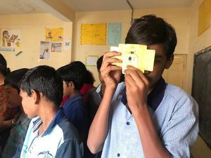
Students with their Foldscopes Thanks to the volunteers, teachers and students of TSWREIS.
Cheers!
Debashree, Ashalatha, Anu, Chandrika and Jayashree
Cheers!
Debashree, Ashalatha, Anu, Chandrika and Jayashree
Sign in to commentNobody has commented yet... Share your thoughts with the author and start the discussion!

 0 Applause
 0 Comments