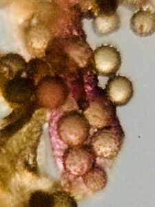
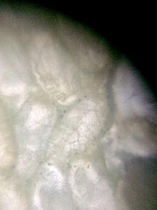
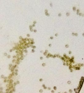
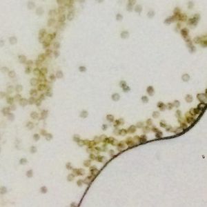
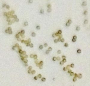
Peristomial teeth and spores of Funaria (moss) under foldscope
 Feb 12, 2019 • 7:57 AM UTC
 Unknown Location
 140x Magnification
 Microorganisms
Kanwar Singh
Learn about the author...
9posts
1comments
1locations
View in Media Gallery
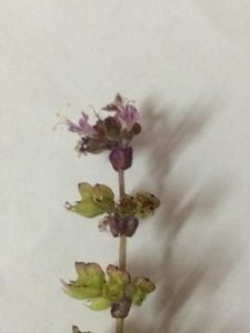
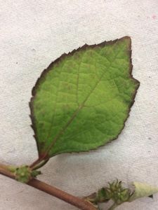
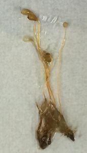
In my practical class of Plant Biodiversity, we were given Funaria plants with sporophyte for study. The sporophyte of Funaria was found to be differentiated into foot, seta and capsule.
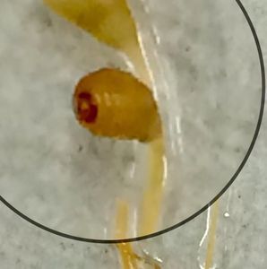
Foot was embedded in the plant body. Seta is a long, cord-like hygroscopic structure that holds the capsule high. Capsule is erect when young but becomes pendant at maturity. Capsule is a pear-shaped structure, the upper portion of which is covered by a cap-like region the operculum, which helps in the dehiscence of capsule and dispersal of spores.
Foot was embedded in the plant body. Seta is a long, cord-like hygroscopic structure that holds the capsule high. Capsule is erect when young but becomes pendant at maturity. Capsule is a pear-shaped structure, the upper portion of which is covered by a cap-like region the operculum, which helps in the dehiscence of capsule and dispersal of spores.
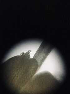
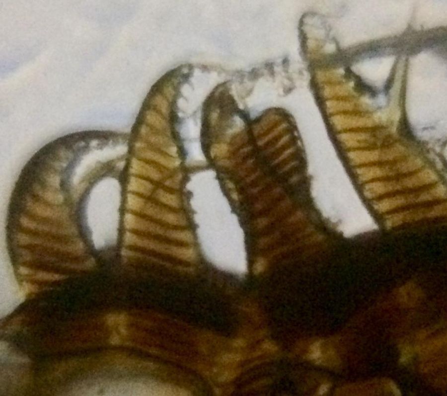
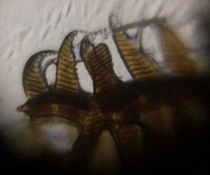
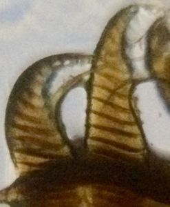
On removing the cap-like operculum from the capsule, teeth-like projections were seen at the top of the capsule. These are peristomial teeth.
These were seen under a foldscope and were seen to be long, conical and present in two rows and in each row there are 16 teeth. Peristome teeth act as a valve that allows the spores to exit the capsule, thus helping in spore dispersal in dry conditions.
These were seen under a foldscope and were seen to be long, conical and present in two rows and in each row there are 16 teeth. Peristome teeth act as a valve that allows the spores to exit the capsule, thus helping in spore dispersal in dry conditions.

Sign in to commentNobody has commented yet... Share your thoughts with the author and start the discussion!

 0 Applause
 0 Comments