Rheinberg illumination for Foldscope
 Feb 18, 2019 • 10:12 AM UTC
 Unknown Location
 140x Magnification
 Microorganisms
Laks Iyer
Human observer of life. https://sukshmadarshin.wordpress.com
97posts
1255comments
5locations
View in Media Gallery
123 years ago Julius Rheinberg invented a variation of the darkfield microscope by adding color to it. Essentially it involved using various combinations of transparent colored papers set up in a way where diffracted light would be of one color and the direct light falling on the object of another. Typically annular rings are made with the annular ring of one color and the central stop of another. You can also make a multi-colored annular ring and get really fancy effects. Read more about those in the following links.
http://www.microscopy-uk.org.uk/mag/indexmag.html?http://www.microscopy-uk.org.uk/mag/artsep00/chillumin.html
https://www.olympus-lifescience.com/ru/microscope-resource/primer/techniques/rheinberg/
The foldscope can also be hacked for Rheinberg illumination and here is how you do it. First, you need transparent films of various colors. I bought mine on amazon, but I remember buying these as a kid at throwaway prices in India. Then you need a desk-lamp on which you can place these papers. Being lazy I didnt make annular rings but just cut them into rectangles. I am sure we can a make a variety of annular ring filters too. So here is how I set it up.
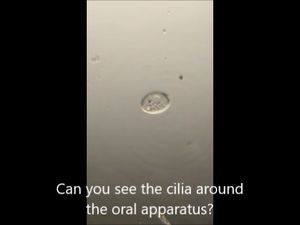
Rheinberg illumination for Foldscope This kind of optical staining has a range of possibilities, but they work best for near transparent life like the optical staining of Amoeba proteus (one of my favorite microscopic organisms). See the next video.
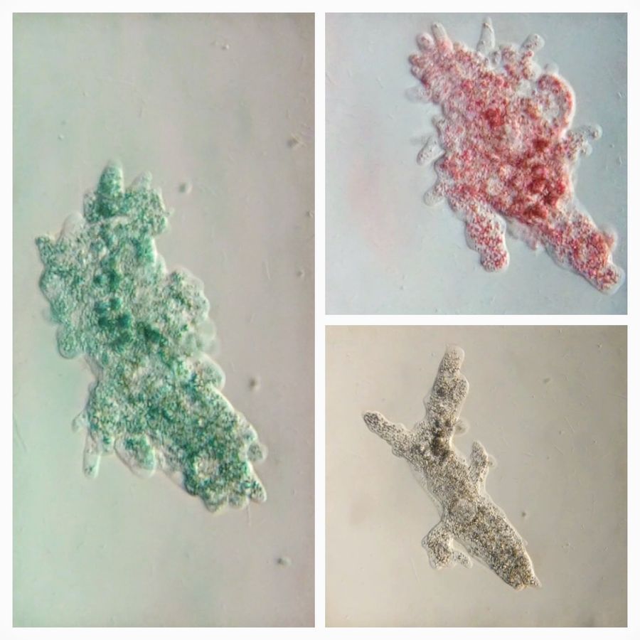
The range of microscopic objects you can enhance by optical staining are left to one’s imagination. In fact I am doing this more than often for everything I see. Some excerpts follow of the organisms I plan to write about soon.
Sign in to commentNobody has commented yet... Share your thoughts with the author and start the discussion!

 0 Applause
 0 Comments
_300x300.jpeg)