DC Micronauts (2016). Foldscoop. 1:1
 Feb 12, 2016 • 4:02 AM UTC
 Unknown Location
 140x Magnification
 Microorganisms
Laks Iyer
Human observer of life. https://sukshmadarshin.wordpress.com
97posts
1255comments
5locations
View in Media Gallery
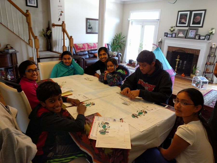
One of the things that Manu and I spoke about after our workshop experiences was to explore the microcosmos with a small group of foldscopers. A few of us in the DC area organized a local foldscope club of about 12 individuals, aptly called the “DC micronauts” (thanks for the name Manu). We range in age from 6-45 years with a mode of 13. Our first meeting was on the 31 st of Jan 2016 in one of our homes. Below is the (Fold)scoop from three of the micronauts describing the day-long event. Pictures and videos were collated from everyone. — Laks
View in Media Gallery
Young micronauts “We used the foldscope to look at many different things like human cells and plant cells. It was interesting to see what plants really looked like up close, but instead of having to use a big microscope, you can simply use a piece of paper- the foldscope. It is also easy to use and assemble. At the beginning the foldscope was just a piece of paper, and a pack of magnets, and a light module. But after some cutting, and folding you hold a fully functional microscope. “
View in Media Gallery
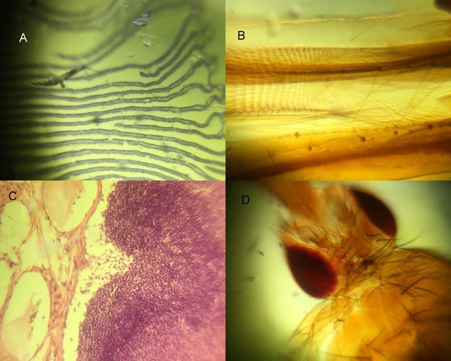
A: Mite eggs on a Jasmine plant, B and C: RBCs under high power, D, plant cell cross-section. — Nina Raghavan- 6 th Grade
“Reflections of a Novice Microbiologist – My First Time Using the Foldscope
Although it was my first time using the Foldscope, I could not have been more excited or impressed at what I discovered using nothing but a paper microscope.
The meeting started off with assembly of the microscope. I had already had mine previously assembled, but received an opportunity to practice assembly again, which I appreciated since I’ll need this knowledge if I am to become a Foldscope expert and educate others on using it properly.
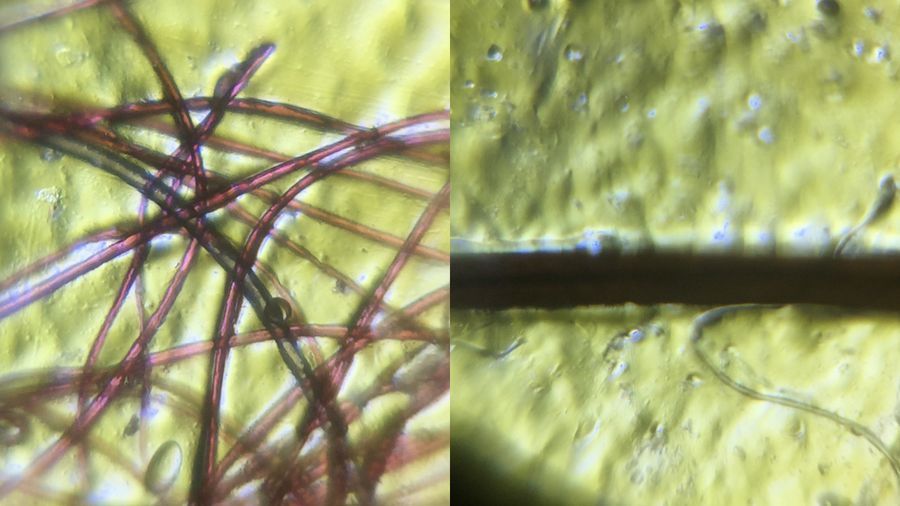
We wasted no time after assembly, and before long all members of the club were peering into their microscopes for the first time. Lax Uncle encouraged us to start off simple, with hair or fibers. I decided to pull out a few fibers from my purple dress and my sister plucked out a strand of her hair, and was enthused to see them up close clearly
“Reflections of a Novice Microbiologist – My First Time Using the Foldscope
Although it was my first time using the Foldscope, I could not have been more excited or impressed at what I discovered using nothing but a paper microscope.
The meeting started off with assembly of the microscope. I had already had mine previously assembled, but received an opportunity to practice assembly again, which I appreciated since I’ll need this knowledge if I am to become a Foldscope expert and educate others on using it properly.
We wasted no time after assembly, and before long all members of the club were peering into their microscopes for the first time. Lax Uncle encouraged us to start off simple, with hair or fibers. I decided to pull out a few fibers from my purple dress and my sister plucked out a strand of her hair, and was enthused to see them up close clearly
View in Media Gallery
Fiber on left, hair on right Soon after, we experimented with a variety of pre-prepared samples to practice using the microscope and camera steadily; out of all my favorite was the insect!
View in Media Gallery
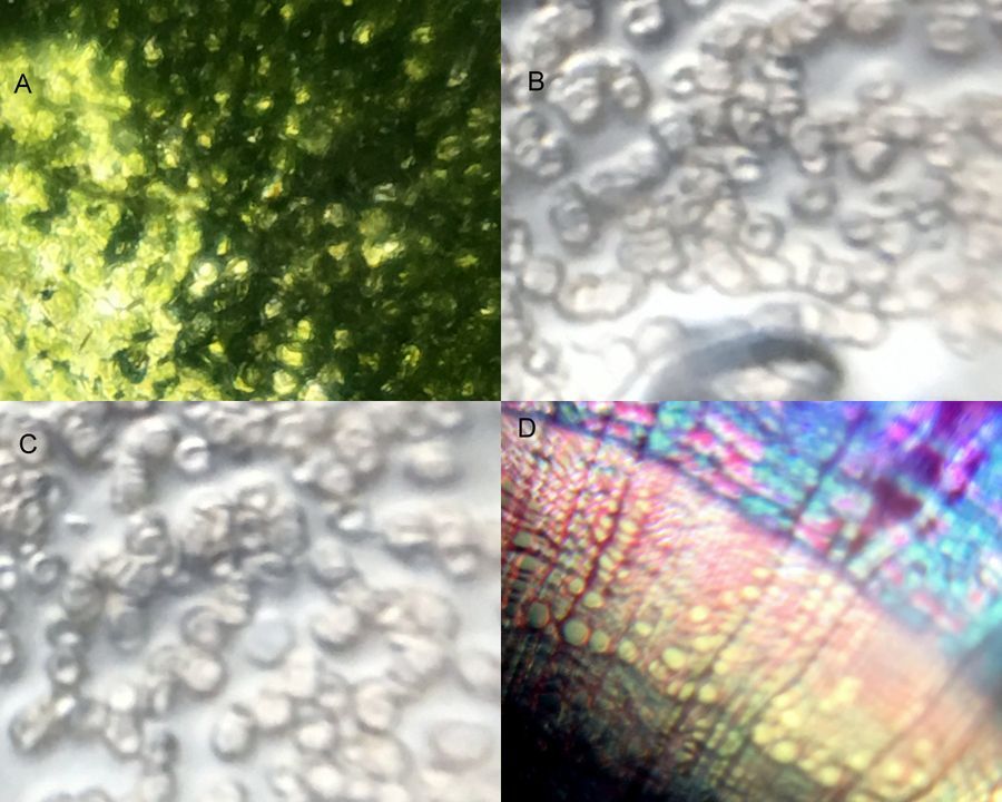
A: Coprinus mushroom, B Housefly part, C, Rabbit lymph node, D. Drosophila head, However, the best part was when we moved on from pre-prepared slides and started experimenting with our own slides. Upon inspection of one the house plants, we were able to discover a mite. It was exciting to witness the mite as it moved around inside the plant, and we used this as an opportunity to take careful observations, starting with the number of legs, (8). The mite also seemed to have two protruding mouth pinchers, called chelisera. Both ticks and mites, which are related, use these to pierce. While ticks use their chelisera to piece skin and drink blood, mites use chelicera to pierce the plant cells and drink the plant’s sap, which I found fascinating.
I also found it amazing to see red blood cells under a microscope for the first time. Lax Uncle explained that while his blood cells were circular shaped, there were cases where people had sickle-shaped blood cells instead. These particular people were affected with a disease commonly known as Sickle Cell Anemia, which originated in Africa but was passed on through generations.
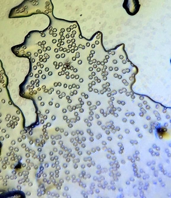
View in Media Gallery
Red Blood cells under Foldscope I was sad to see the meeting come to the end, but what I had discovered and experienced got me thinking about something more.
If I had been able to learn so much about the endless world under the microscope in a few hours, then the possibilities for the future were endless. Should I continue to experiment, wonder, and question the world around me inside and outside the club, I’ll not only grow as a scientist and microbiologist, but also a creator, a thinker, a dreamer; someone who has the possibility to discover new phenomena and change the world. What I’ve been able to witness is just the beginning.”— Reethi Padmanabhan 9 th grade .
”

View in Media Gallery
First two photos: Nymphae of Acquisto stem, third: Apple skin ”
–Pics from Kartik Krishnan 11th grade.
It of course ended with food and fanfare. Foldscoping makes your hungry :). The micronauts have already made a few individual posts and seemed to have taken to it quite seriously. Keep your eyes open for more posts from us and looking forward to your feedback and ideas. Hope to see other such foldscope clubs across the globe.- DC Micronauts
Aditi, Yash, Nataraj, Kartik, Uma, Nina, Vaishnavi, Reethi, Keerthi, Shivaprasad, Aruna, Laks
Sign in to commentNobody has commented yet... Share your thoughts with the author and start the discussion!

 0 Applause
 0 Comments
_300x300.jpeg)


