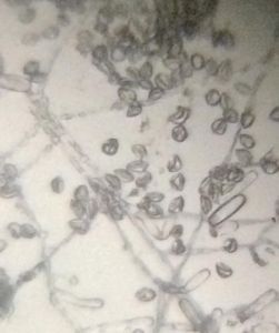
Powdery mildew on Tulsi under foldscope
 Mar 03, 2019 • 8:32 AM UTC
 Unknown Location
 140x Magnification
 Microorganisms
Samriti Dhawan
Learn about the author...
9posts
0comments
1locations
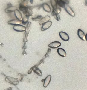
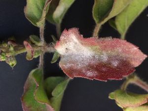
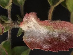
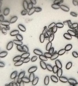
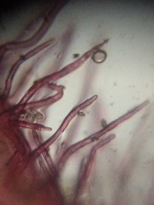
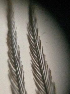
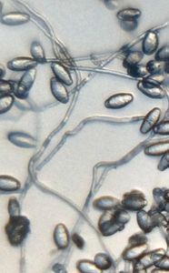
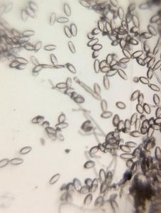
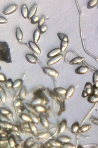
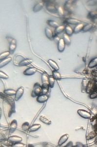
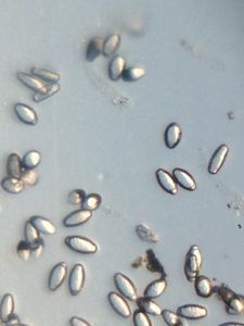
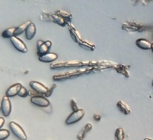
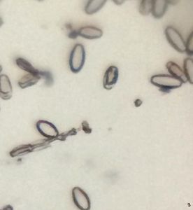
White powder was seen on upper side of some tulsi leaves. Also the leaves showing powdery mass had turned reddish-brown in colour. The powdery mass when seen through foldscope showed large number of microscopic fungal spores – probably conidia. On the conidiophores, the point of attachment of conidia were clearly marked. Besides, the spores were oval in shape, with double layered wall and a plug-like struture at the tip.
Sign in to commentNobody has commented yet... Share your thoughts with the author and start the discussion!
 0 Applause
 0 Comments