The hunt for white blood cells..
 Mar 13, 2016 • 9:41 PM UTC
 Unknown Location
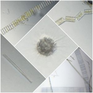
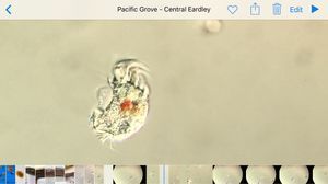
 140x Magnification
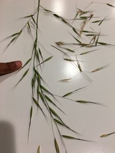
 Plants
Saad Bhamla
Learn about the author...
32posts
11comments
2locations
View in Media Gallery
This is a second part of exploring what lies in our bodies using the foldscope. Apart from the obvious utility in seeing what we’re made off, I also think this is a useful collection of experiments that you can use at any place or time for a quick foldscope demonstration. The last experiment was with cheek cells. This post is on looking at blood and hunting for white blood cells…
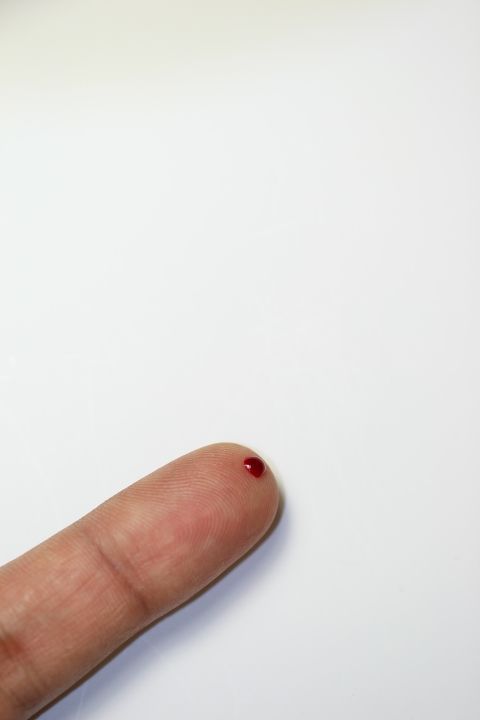
The first step is to use a sterile lancet on your finger and generate a tiny droplet of blood (proceed with caution at this step and conduct this step under adult supervision).
The first step is to use a sterile lancet on your finger and generate a tiny droplet of blood (proceed with caution at this step and conduct this step under adult supervision).
View in Media Gallery
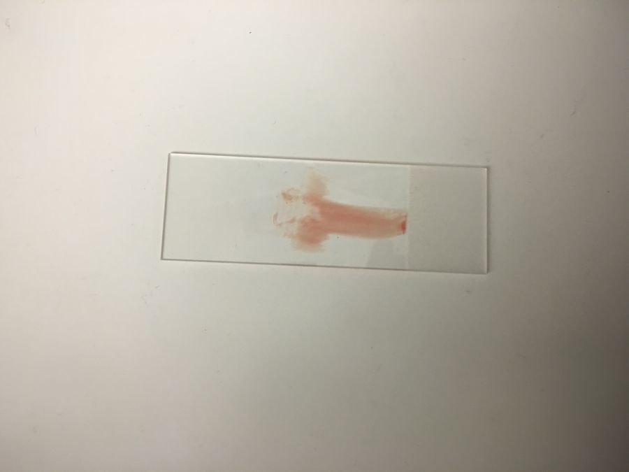
The next step is to transfer the blood onto a clean slide and create a smear. See video below.
I put a cover-slip on the slide and this is how the slide looked before mounting onto the foldscope.
View in Media Gallery
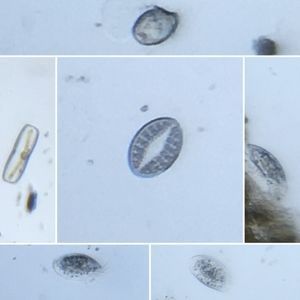
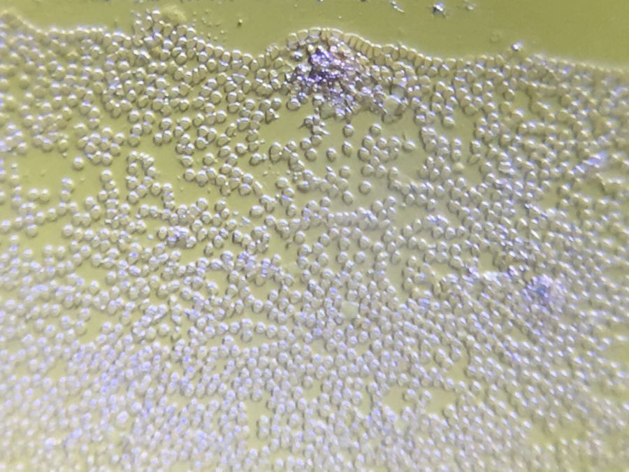
Under the foldscope, you see hundreds and thousands of red blood cells (RBC’s). It’s amazing how many of them are in a teeny tiny drop of blood.
Now, we wanted to hunt for white blood cells – these are fewer in number and thus harder to find. One trick we used next is to stain them with Methylene blue solution. The idea is that the methylene blue dye would stain the cytoplasmic material in the white blood cells, and they would appear blue amidst a sea of red blood cells. This is akin to a game of ‘Where’s Waldo’ – and we use a trick to color Waldo blue so it becomes trivial to locate him..
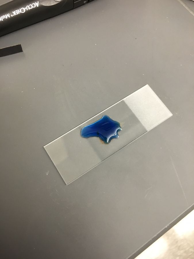
The way we did it is to first fix the cells with methanol (you could potentially use ethanol from a local drugstore too): after creating a smear, we air-dried it for a few minutes, and then added a few drops of methanol and let it evaporate (2-3mins). Once the methanol had evaporated, we added a few drops of methylene blue (you can find this online easily). We let it sit for about 5 mins. Then washed the slide off under tap-water.
The way we did it is to first fix the cells with methanol (you could potentially use ethanol from a local drugstore too): after creating a smear, we air-dried it for a few minutes, and then added a few drops of methanol and let it evaporate (2-3mins). Once the methanol had evaporated, we added a few drops of methylene blue (you can find this online easily). We let it sit for about 5 mins. Then washed the slide off under tap-water.
View in Media Gallery
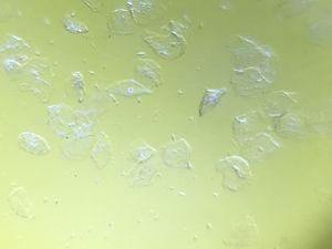
Blood smear stain with Methylene blue
View in Media Gallery
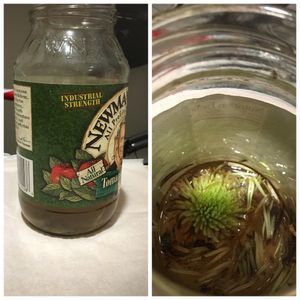
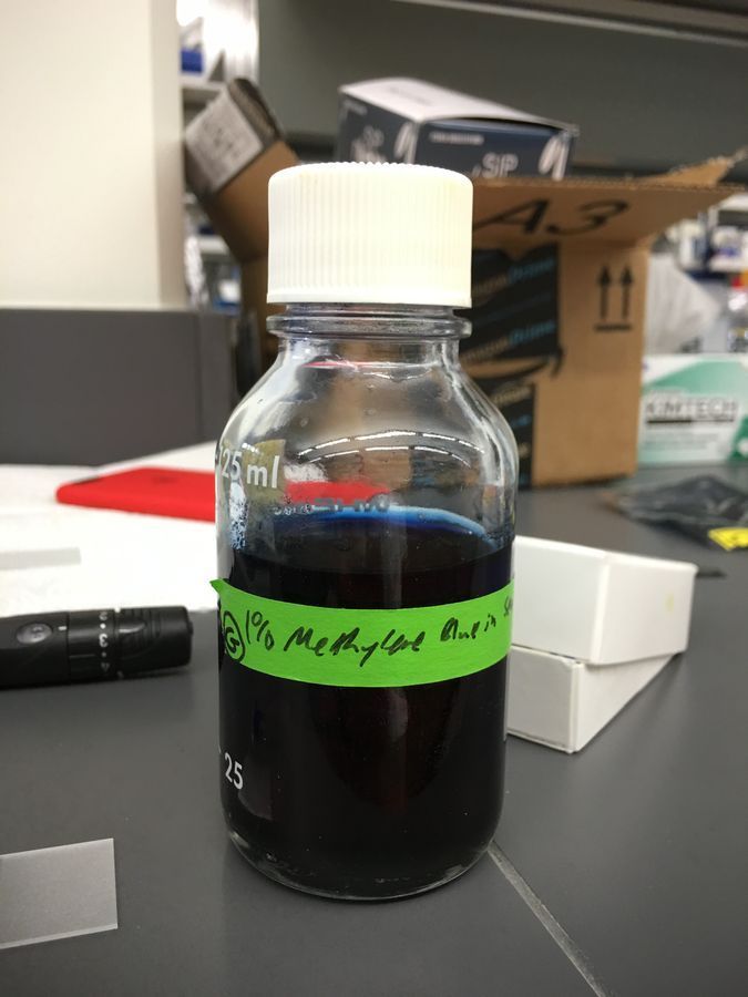
1% Methylene blue in bottle
View in Media Gallery
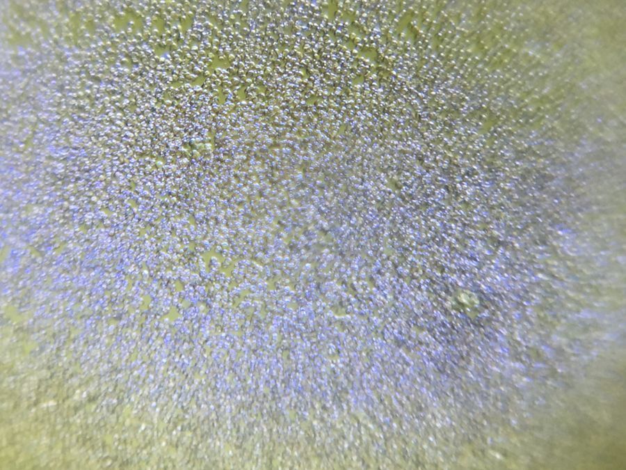
red blood cells
View in Media Gallery
white blood cell with stain After covering the smear with a cover-slip, we mounted it under a foldscope. In the next video, in the first few frames, we believe we saw a stained white blood cell in the top-left corner.
One thing became apparent very quickly that there are far more red blood cells than white blood cells – and we only could see some bluish shapes here and there – that is why this post is titled a hunt for white blood cells – they are elusive and tricky to find at a first-try.
Here’s another video where we scan the smear – take a look.
Here’s another video where we scan the smear – take a look.
This excercise imparted a few thoughts on my mind. For disease diagnostics, looking at a smear under a microscope can be challenging if you are looking for white blood cells or parasites. Hence staining well is a very useful trick to know, especially if one has to eliminate false-negatives. This also impresses the importance of centrifugation to remove the RBC’s from the picture – to really be able to separate out the objects of interest from the red blood cells.
Hopefully, if you are out in the field, in a class-room or at home demoing the foldscope, in addition to cheek cells, now you have a second easy sample to quickly prepare and share with others, the wonders of the micro-cosmos of you own body.
DOI: 10.15200/winn.145806.66635 provided by The Winnower , a DIY scholarly publishing platform
Sign in to commentNobody has commented yet... Share your thoughts with the author and start the discussion!

 0 Applause
 0 Comments