Tepal epidermis
 Mar 18, 2016 • 4:54 AM UTC
 Unknown Location
 140x Magnification
 Microorganisms
Laks Iyer
Human observer of life. https://sukshmadarshin.wordpress.com
97posts
1255comments
5locations

View in Media Gallery
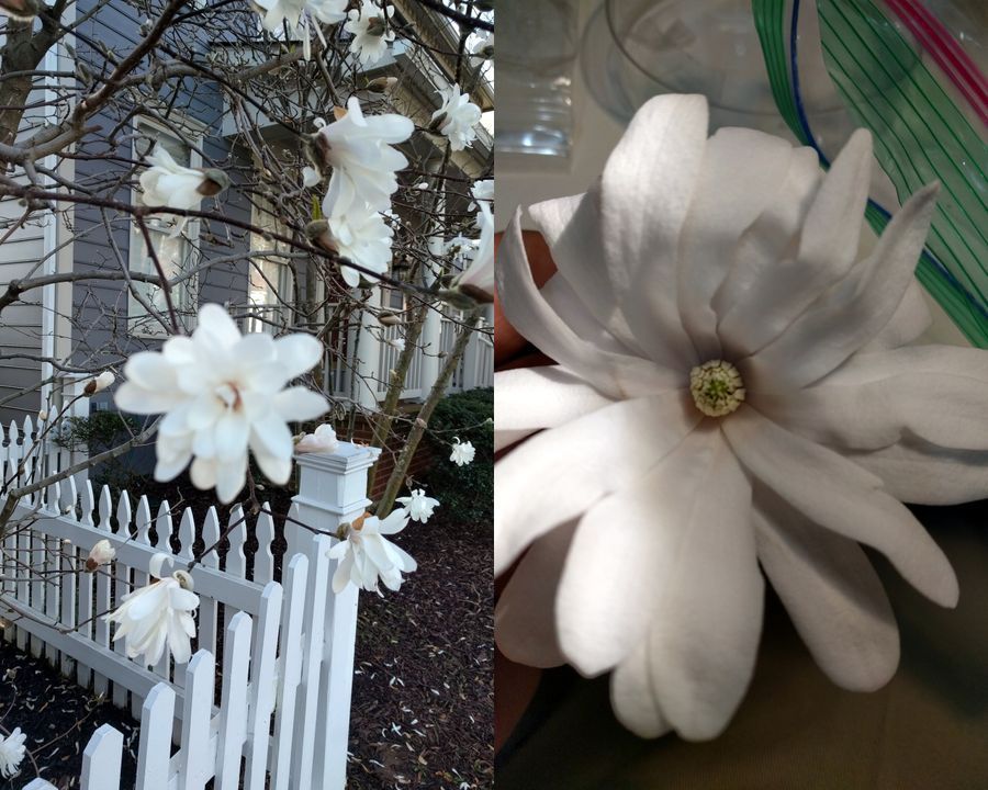
You might have seen this earlier post by Cristina on plant histology . I loved the jigsaw pattern on the leaf epidermis and wanted to see epidermal patterns in other plants and their structures. One of the flowers that one sees in early spring in the DC area are the magnolias. This one in the picture below is Magnolia stellate, a native of Japan with a sweet smell and over 20 tepals (petals and sepals are fused in these and called so). The tepal is thick and somewhat rubbery.
View in Media Gallery
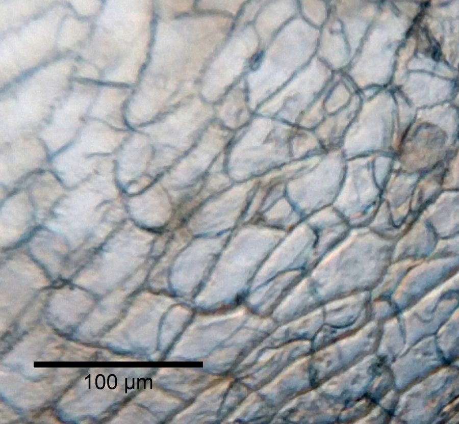
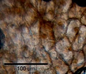
However, I realized that peeling the epidermis of this tepal is rather easy. Just break the tepal to the point it is almost in two and pull one over the other to peel the epidermis. I did this for the upper and lower epidermis. I also stained it with Carbol Rose Bengal and could see really nice nuclei. The epidermises dont seem very different from each other. I wonder what is the range of epidermal shapes?
Click on images below for hires pictures
Source Unstained Stained Petal Upper epidermis
Click on images below for hires pictures
Source Unstained Stained Petal Upper epidermis
View in Media Gallery

View in Media Gallery
Petal lower epidermis

Sign in to commentNobody has commented yet... Share your thoughts with the author and start the discussion!

 0 Applause
 0 Comments
_300x300.jpeg)


