Apr 15, 2019 • 8:41 PM UTC
 Unknown Location
 140x Magnification
 Unknown
Meena Hari
Learn about the author...
9posts
0comments
2locations
View in Media Gallery
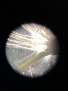
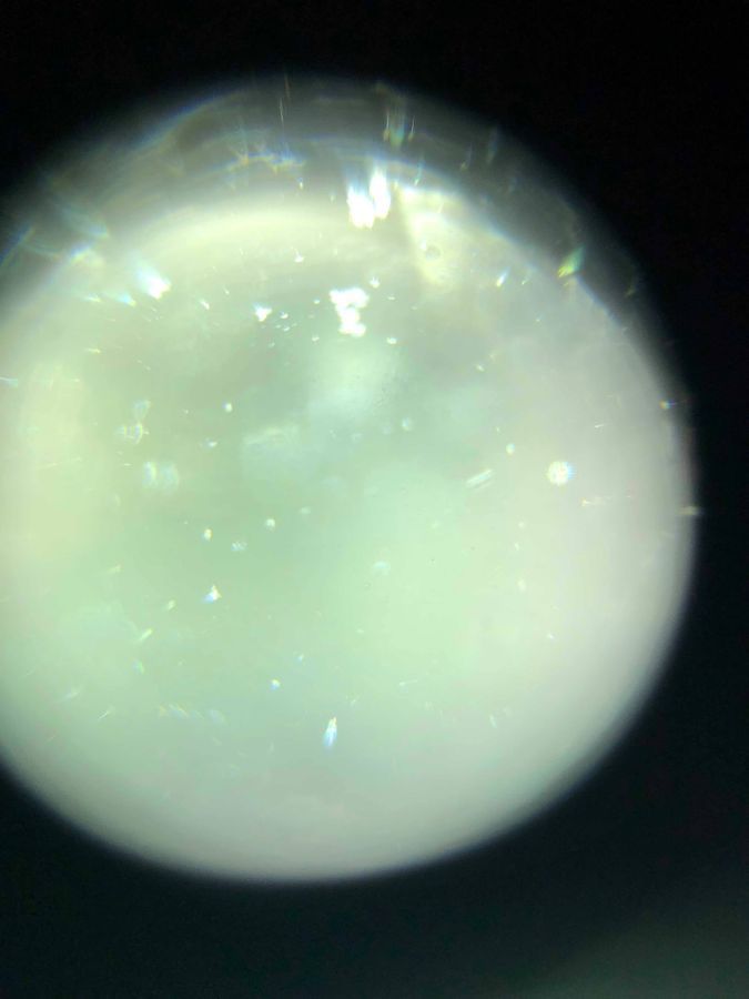
This image is a magnification of water from Millikan pond. It was obtained midday on 4/9/19. The water sample was obtained by filling a plastic cup of water with pond water (dragging it across the pond surface). The domain of the spiky organism in the center, right of the image is likely Bacteria. I believe this because it seems to resemble the shape of a coccus virus bacteria. Eukarya, for example, on the other hand, would likely appear more complex (have more organelles, while the microorganism in the sample looks quite simple). The foldscope has a magnification of 140x and the bacteria could be roughly 1 micrometer.
1) I wonder whether the microorganism is truly a bacteria/archaea/life-form or just some type of debris. We could try Gram staining the sample or culturing the bacteria to see what kind of bacteria it is/whether it really is a living microorganism.
2) I also wonder how much of the integrity of the sample is maintained under magnification with the foldscope because I found that my foldscope’s lens itself had particles on it (without the sample present) that I was unable to clean. Here, we could use a different foldscope or lens to view the sample and verify which parts of the image are truly part of the sample and which are not.
Apart from the spiky organism observed above, much of the rest of the image seems to be just water or debris.
#caltechbi1
Sign in to commentNobody has commented yet... Share your thoughts with the author and start the discussion!
 0 Applause
 0 Comments