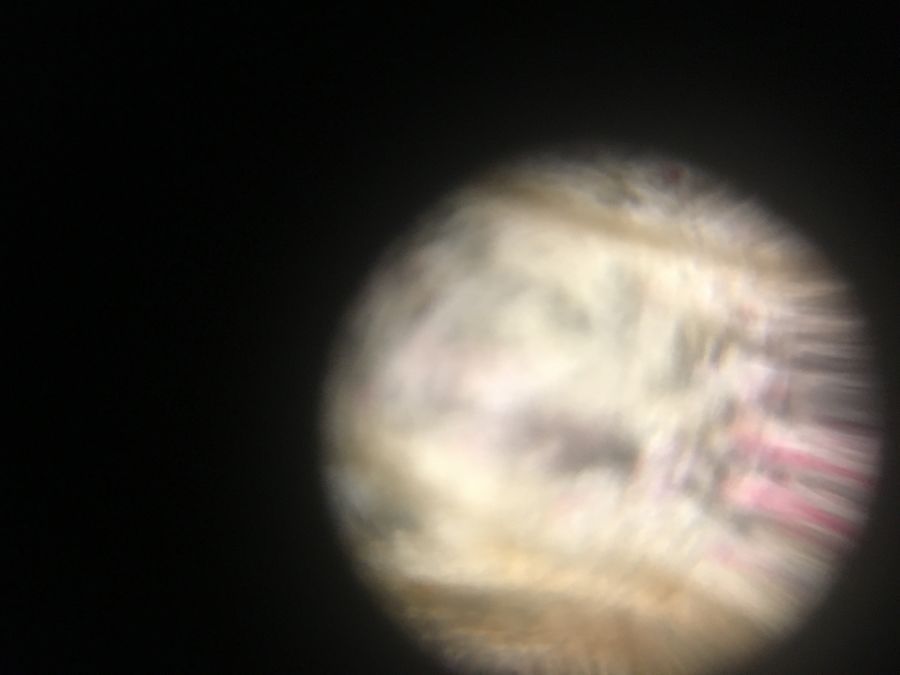
Pink flower petal at 140x
 Apr 17, 2019 • 10:47 AM UTC
 Unknown Location
 140x Magnification
 Unknown
Rashida Hakim
Learn about the author...
2posts
0comments
2locations
View in Media Gallery
This photo shows the cells and veins in a flower petal collected from Caltech campus on 4/10/15 at 6 pm . The sample was removed from a living flower and then pressed between 2 clear stickers until the petal itself turned translucent (approximately 5 minutes). We can see the pink pigment in some of the cells of the petal. One question I have about my image is: what shape are the cells in the petal? In this image they appear to be thin rectangles but we could find out more information by taking pictures of a different cross-section of the petal. Another question I have is what is being transported in the veins of the petal currently? Answering this question would likely require a microscope with significantly better magnification so the molecules in the veins could be observed. #caltechbi1
Sign in to commentNobody has commented yet... Share your thoughts with the author and start the discussion!
 0 Applause
 0 Comments