Live Mealworms eating plastic
 Apr 19, 2016 • 11:59 AM UTC
 Unknown Location
 140x Magnification
 Microorganisms
Saad Bhamla
Learn about the author...
32posts
11comments
2locations

View in Media Gallery
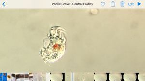
This is probably the most bizarre and coolest organism I’ve ever seen under a foldscope. It’s a mealworm that eats Styrofoam (and other plastic) and degrades into biodegradable waste.
I’d read about these worms on the Stanford website, so when the researchers (Wei-min, Craig , Anja and Shandhan) who discovered them invited us to take a look at these worms using the foldscope, I jumped at the opportunity.
Before we jump into the videos, I have to share that the researchers were looking at these worms live under a microscope for the first time, and you can hear their excitement in their voices. The videos are also super long, but I hope you will enjoy the discovery journey we made together, and saw for the first time the sharp jaws of these worms, them munching in action, their negative reaction to light, and their gut movements.
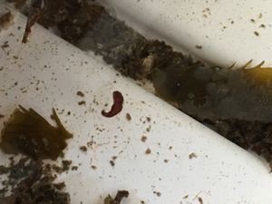
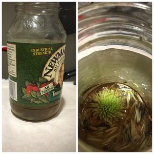
So here’s a first video using an iPhone of the meal worms in a plastic container, enjoying their styrofoam snack. This should give you a sense of scale (upto 1cm long).
I’d read about these worms on the Stanford website, so when the researchers (Wei-min, Craig , Anja and Shandhan) who discovered them invited us to take a look at these worms using the foldscope, I jumped at the opportunity.
Before we jump into the videos, I have to share that the researchers were looking at these worms live under a microscope for the first time, and you can hear their excitement in their voices. The videos are also super long, but I hope you will enjoy the discovery journey we made together, and saw for the first time the sharp jaws of these worms, them munching in action, their negative reaction to light, and their gut movements.
So here’s a first video using an iPhone of the meal worms in a plastic container, enjoying their styrofoam snack. This should give you a sense of scale (upto 1cm long).


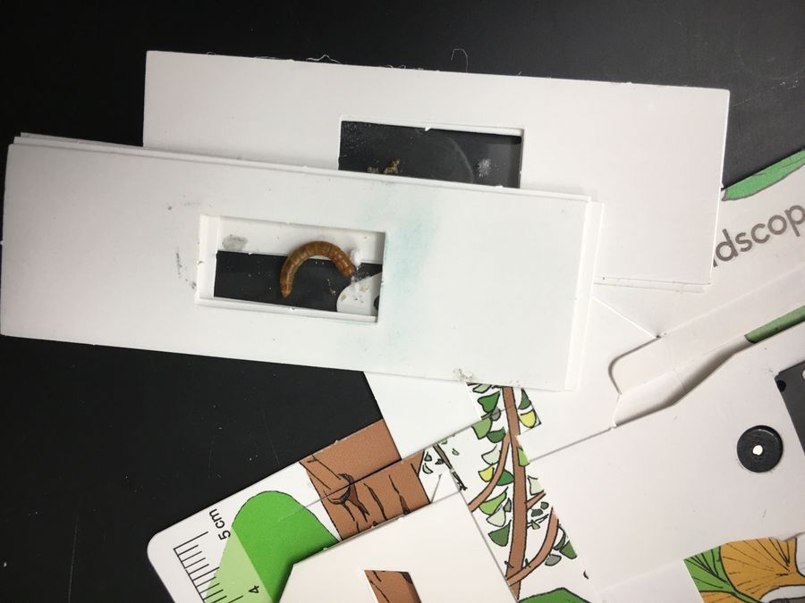
We first took a small mealworm (early instar) to ensure we could see them under a foldscope. We mounted them by creating a ‘jail’ using paper slides and clear-tape.
View in Media Gallery
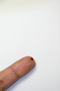
The next image gives a comparison between the size difference between larvae. We first mounted the smaller one followed by the larger one (on right).

View in Media Gallery
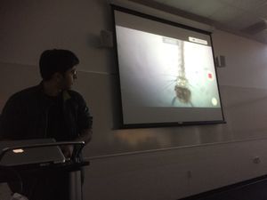
Ok. So this is the first video. Keep a ear open for the excitement of the researchers who are seeing their organism live under a microscope for the first time.
Couple of points to observe from this first video:
Sharp claws. Jim has really good ideas. I had to go back and re watch the video to really see the claws clearly. They are quite sharp. Why do they need such sharp claws? Squishy legs. Quite unusual. 3 pairs of legs and 1 pair of antennae. We mistakenly count antennae as legs. The discharge event- that’s how it ejects the digested plastic. Researchers can look closely at it to quantify the efficiency of plastic degradation. I understand that not all the styrofoam is digested- perhaps there are micro particles in the poop? Counting the thorax segments can help us identify the developmental stage.
Sharp claws. Jim has really good ideas. I had to go back and re watch the video to really see the claws clearly. They are quite sharp. Why do they need such sharp claws? Squishy legs. Quite unusual. 3 pairs of legs and 1 pair of antennae. We mistakenly count antennae as legs. The discharge event- that’s how it ejects the digested plastic. Researchers can look closely at it to quantify the efficiency of plastic degradation. I understand that not all the styrofoam is digested- perhaps there are micro particles in the poop? Counting the thorax segments can help us identify the developmental stage.


Okay. So now we wanted to see its sharp jaws and mandibles that it uses to chew on plastic, so we flipped the slides over..
There is a big WHOA moment when we first catch sight of the jaws (4:40). Wow. What a moment.

View in Media Gallery
Now we decided to see if we could see in its gut and actually see plastic particles. The next video wasn’t successful but you can see gut movement


In the next video, you actually see contractions of the gut due to transmission of light from the back-side. Keep an eye out for the neurons. These are sensitive to light and make the larvae phobic to light, which will become apparent in the next video. I imagine the neurons protect the larvae from excess light-exposure which can be harmful.
This is the last and final video. It is full of wonderful serendipitous moments. Watch it first and I’ll list out a few below for your reference. We actually have a piece of styrofoam in the slide to see if we could catch the worm in ‘action’. Watch to find out..
We see the worm bite on the paper slides and start chewing. Do you see how it responds to agitation by light? Can you see how it’s using its leg and claws to hold on?

View in Media Gallery
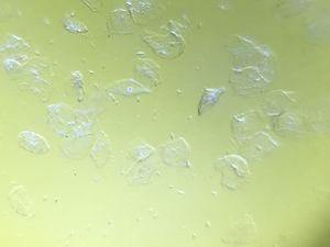
Here’s an image of the worm and the holes it made on the slide.

View in Media Gallery
Hope you enjoyed this post. Special thanks to Wei-min, Craig , Anja and Shandhan for sharing the worms. And Manu for providing the opportunity.
Keep exploring,
Saad
This post is open to read and review on The Winnower.
Keep exploring,
Saad
This post is open to read and review on The Winnower.
Sign in to commentNobody has commented yet... Share your thoughts with the author and start the discussion!

 0 Applause
 0 Comments