10 Samples for Caltech Bi1
 May 29, 2019 • 8:48 PM UTC
 Unknown Location
 140x Magnification
 Unknown
Katerina Gorou
Learn about the author...
3posts
0comments
1locations

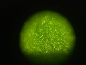
In this image, the cells on the top outer surface of a basil leaf can be seen. A bright flashlight was used to light the sample, making the cells seem glassy and bright. The sample was obtained at 11:40 am on 5/9/19 at my home in Forsyth, IL from a basil plant. The sample was obtained by tearing a small leaf off of the plant.
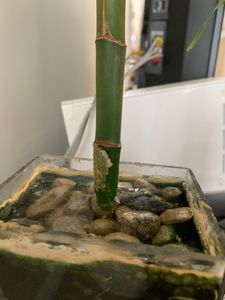
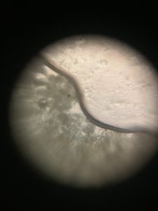
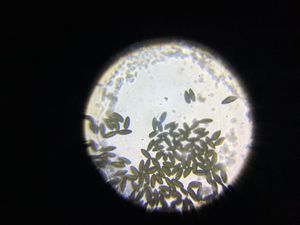
In this image, a dark, wormy-looking microorganism can be seen. This organism was found on a lucky bamboo plant that had grown some white crust. I wonder what could’ve caused the plant to accumulate all this off-white crust, because it did not look like this last break. The sample was obtained at 12:30 pm on 5/9/19 at my home in Forsyth, IL from the crusty white material on the stem of a lucky bamboo plant. The sample was obtained using a toothpick to scrape it onto the slide.


In this image, some red and pretty large blood cells from my right pinky can be seen. I initially assumed they were red blood cells, since they were red, but the cells in the image all seem to have a nucleus. Although white blood cells are nucleated, none are this big, so these aren’t white blood cells either. I considered that they may just be several RBCs grouped together, but that wouldn’t explain the nuclei seen. One final possible explanation is that these are squamous epithelium cells from my pinky that were stained red by the blood, but I don’t think this is likely either. So, I am unsure of the identity of these cells. This sample was obtained at 6:50 am on 5/10/19 at my home in Forsyth, IL from my right pinky. The sample was obtained by pricking my pinky with a needle and squeezing blood onto the slide.
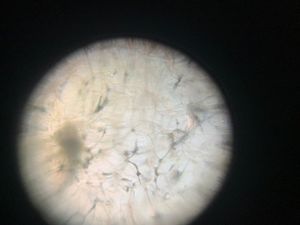
In this image, cells from the inside of a cucumber can be seen. They are all very translucent, separated by faint, wrinkly cell walls. The sample was obtained at 2:55 pm on 5/10/19 at my home in Forsyth, IL from a English cucumber. The sample was obtained by using a knife to cut an extremely thin slice of cucumber, which was placed on the slide.
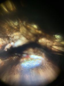
In this image, cells from the cap of a mushroom can be seen. This image is very interesting because there is a variety of colors visible, including a small red patch. Other than that, the image is comprised of brown/black filaments crossing over each other. The sample was obtained at 4:51 pm on 5/28/19 by Dabney House at Caltech. The sample was obtained by ripping off a small portion of the mushroom cap.

In this image, cells from a jelly bean succulent can be seen. Similar to the cucumber cells, the cells are all very translucent and well-defined. There are also small dimples/spots within the cells. The sample was obtained at 5:02 pm on 5/28/19 by Schlinger Laboratory at Caltech. The sample was obtained by cutting off a small piece of the succulent with scissors, and then cutting a thin slice for the slide.
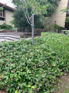
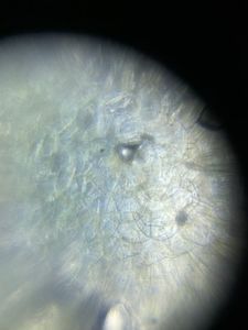
In this image, cells from a jasmine petal can be seen. The small cells don’t seem too well-defined in the center, but are much clearer around the edges. Also, I think some parts look a bit brown because I foldscope’d the sample a few afters after obtaining it. The sample was obtained at 5:03 pm on 5/28/19 by Noyes Laboratory at Caltech. The sample was obtained by tearing off a jasmine flower from the bush, and then placing a petal on the slide.


In this image, cells from a purple flower’s petal can be seen. I find it very interesting that the colors in this petal are in strips, and that not the entire petal is purple (and that part of it is green). The sample was obtained at 4:55 pm on 5/28/19 by the Hameetman Center at Caltech. The sample was obtained by cutting off a small purple bud from the plant, and then tearing off a petal from the bud.
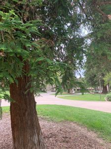
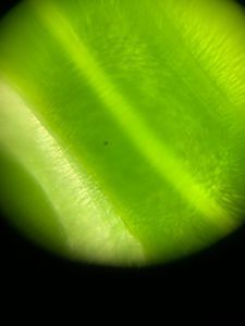
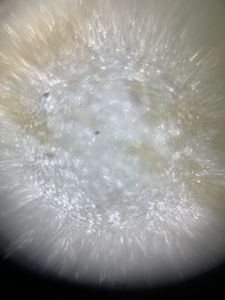
In this image, needles from what I believe is a bald cypress tree can be seen. It seems that the center strip of the green needle is thinner/clearer than the rest of the needle because it is a lighter shade of green. The sample was obtained at 4:57 pm on 5/28/19 by Spalding Laboratory at Caltech. The sample was obtained by tearing off a small bunch of needles from the tree.
View in Media Gallery
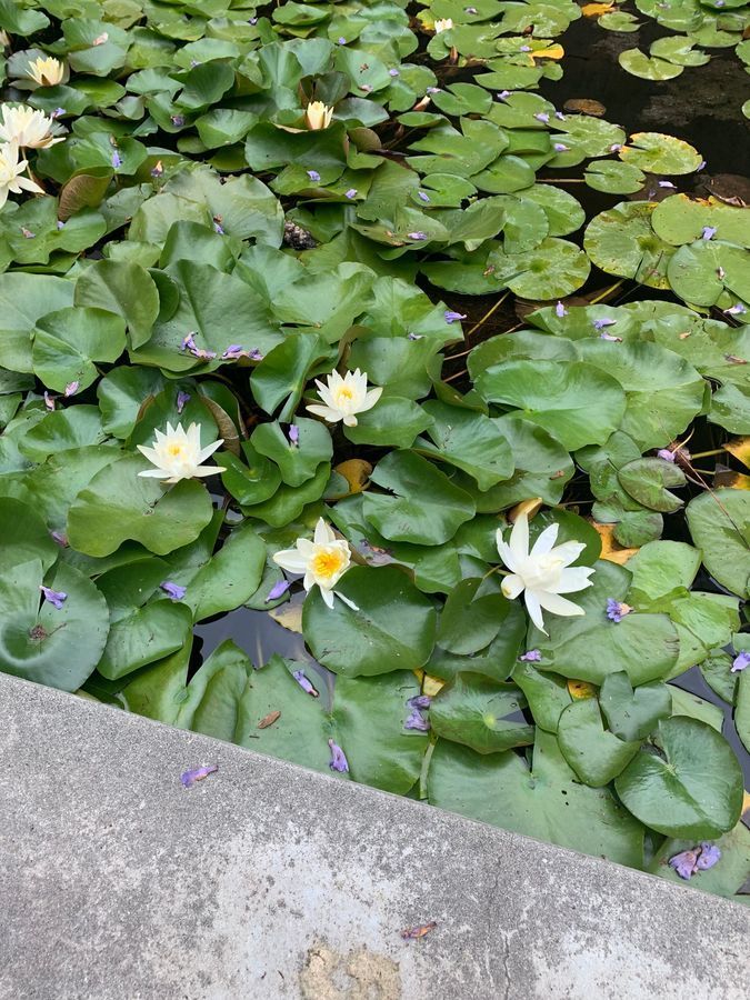
In this image, the cells of from a lily pad’s flower’s petal can be seen. Similar to the jasmine petal cells, the cells are whitish/clear, and there doesn’t seem to be that much to see. The cells look elliptical/circular. The sample was obtained at 3:00 pm on 5/27/19 from the lily pad pond in front of Beckman Laboratory at Caltech. The sample was obtained by tearing off part of a lily pad flower’s petal. #caltechbi1
Sign in to commentNobody has commented yet... Share your thoughts with the author and start the discussion!
 0 Applause
 0 Comments