Fun with Fungi
 May 08, 2016 • 1:46 PM UTC
 United States
 140x Magnification
 Microorganisms
Damon Tighe
I currently work as a Curriculum and Training Specialist at Bio-Rad laboratories and help teachers incorporate more biotechnology into their classroom. I worked in DNA sequencing for years including the Human Genome Project and single cell genomics (termite, cow, and other guts). I have a love of the outdoors that I spread to the public via Calnature.org. I enjoy backpacking, picking mushrooms and photography.
15posts
17comments
9locations

View in Media Gallery
In North America fungi are a mess, taxanomically speaking. Many fungi that look similar to ones found in Europe inherited the same scientific names and this is partly because beyond the fruiting body (mushroom), fungi are pretty hard to get a good look at. A fair number of attributes that are used to discriminate between species need microscopy. DNA sequencing of a conserved region such as the ITS is becoming the gold standard, but its a much harder attribute to access for most amateur mycologist. Access to microscopes for looking at the asci and basidia (sexual reproductive structures) in addition to spore size, shape, and texture could greatly improve observation data on fungi and can help in the identification of new species and/or getting the taxonomy straightened out on mis-labeled species.
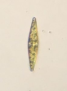
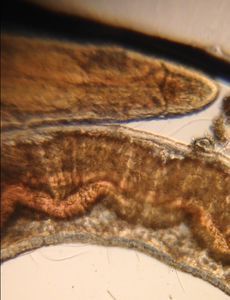
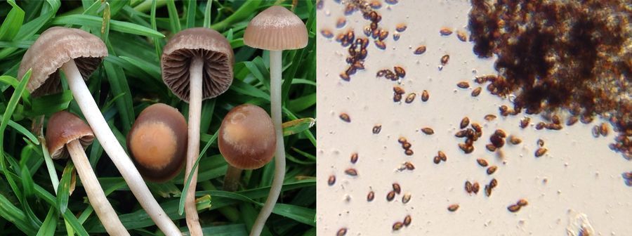
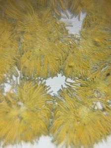
I went for a walk around Lake Merritt in Oakland, California to see if any fungi had sprouted up since the past rain and was happy to find one fresh fruiting of Paneolina foenisecii that I was able to use the foldscope to look at its spores:
I went for a walk around Lake Merritt in Oakland, California to see if any fungi had sprouted up since the past rain and was happy to find one fresh fruiting of Paneolina foenisecii that I was able to use the foldscope to look at its spores:
View in Media Gallery
Paneolina foenisecii and spores (12-14 x 6-7.5 µm from literature)
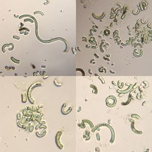
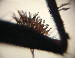
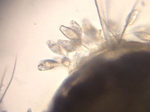
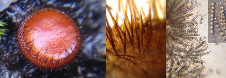
I had some dried mushroom samples from recent hikes, so I re-hydrated them and also looked to see how well Foldscope did at viewing their spores and asci. I specifically looked at the Ascomycota because the spores tend to be 10µm or greater which seems to be ideal for the Foldscope’s low magnification lens. I have yet to master getting good images with the high magnification lens.
I had some dried mushroom samples from recent hikes, so I re-hydrated them and also looked to see how well Foldscope did at viewing their spores and asci. I specifically looked at the Ascomycota because the spores tend to be 10µm or greater which seems to be ideal for the Foldscope’s low magnification lens. I have yet to master getting good images with the high magnification lens.
View in Media Gallery
Scutellinia sp.
View in Media Gallery
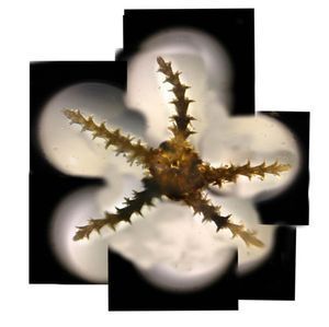
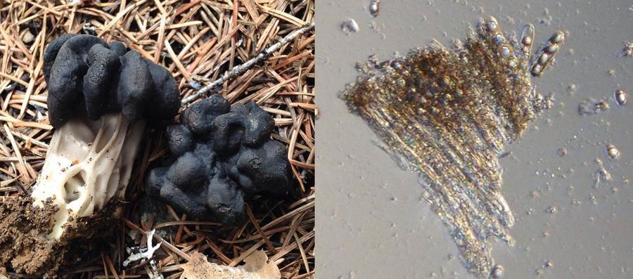
Helvella vespertina
View in Media Gallery
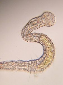
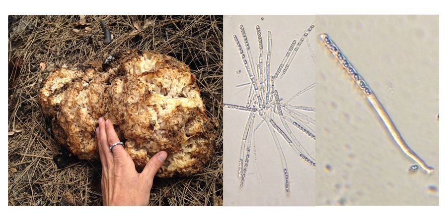
Peziza proteana ssp. sparassoides
Notes on technique: I used a glass slide with a coverslip over a drop of water with the spores/scraped sporing surface. I found that being able to wipe the lens of the Foldscope with some lens paper greatly increased my image quality, but this is probably because I had been getting water with all kinds of stuff in it when I was messing around focusing. I had a desk lamp that I would move the Foldscope in front of until I found the appropriate contrast to get a good image with an iPhone 5. I would zoom in with the cell phone’s camera, push down on the screen to auto-focus lock and then use the onscreen slider to adjust the exposure. Shooting spores was a little difficult because I tend to hold the Foldscope up vertically and the spores start to slide down inside the coverslip and as I focus I occasionally put pressure on the coverslip causing flushes of water to whisk spores away from my field of view.
Overall I’m really happy with Foldscope’s ability to help see these structures in the field as it could help with in field identifications and decisions about what specimens are worth taking out of the field for further study.
Sign in to commentNobody has commented yet... Share your thoughts with the author and start the discussion!
 0 Applause
 0 Comments