What makes tomatoes red?
 Jul 01, 2019 • 12:55 PM UTC
 Unknown Location
 140x Magnification
 Microorganisms
Mitali
Learn about the author...
30posts
93comments
4locations
View in Media Gallery
Hello all,
Did you know, the tomato and cherries get the colour on their skin by different ways ?
Here is our experiment and its immediate replication.
The power of foldscope and social media.
On a rainy Sunday evening.. Deepak’s daughter suddenly decided to do some foldscope experiments.
After a few random stints, they stumbled on tomato. They peeled out the skin and pulp and mounted them separately.
The idea was to see what gives tomato skin its colour.
Expecting the pigment inside the cells, they took some images and voila!
View in Media Gallery
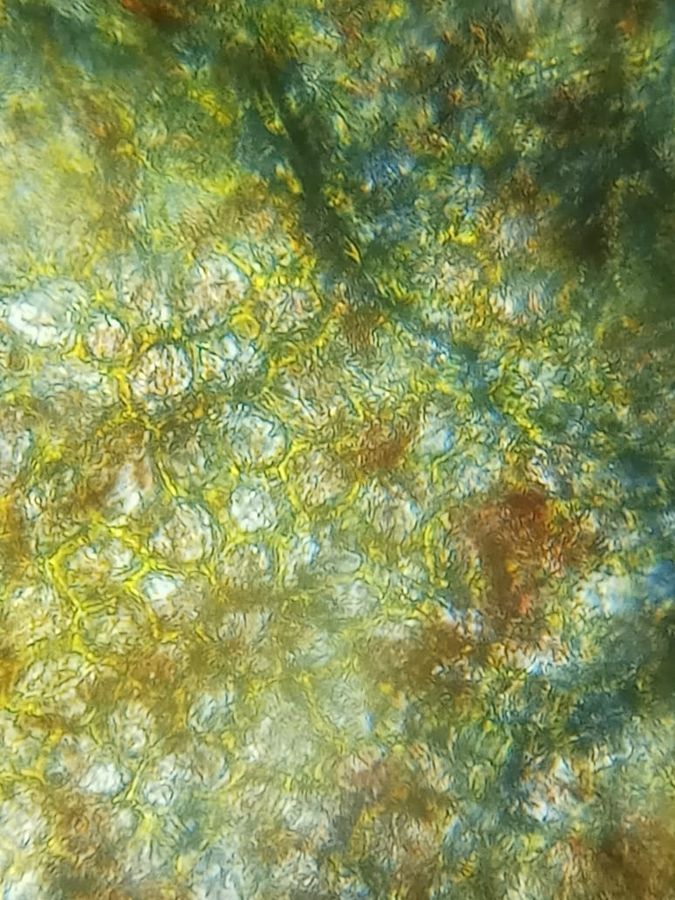
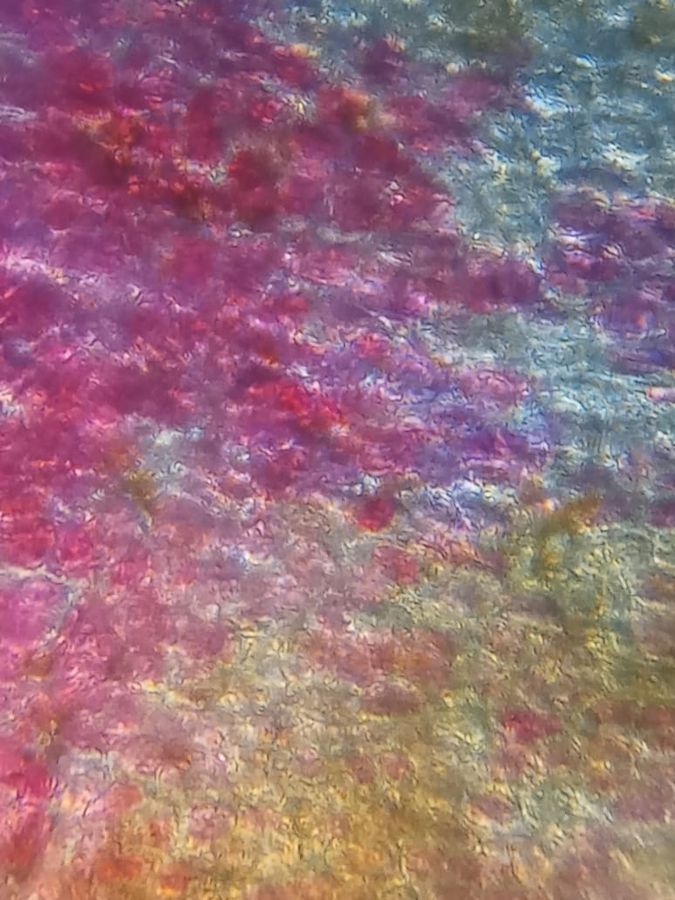
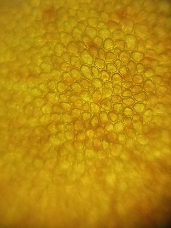
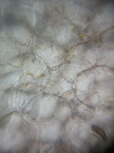
Tomato Peel
The cells were empty and the yellow orange pigment was seeming outside the cells. The cells appeared packed, but empty and the pigment all around it.
More interestingly the cherry skin had pigment inside the cells !!
The cells were empty and the yellow orange pigment was seeming outside the cells. The cells appeared packed, but empty and the pigment all around it.
More interestingly the cherry skin had pigment inside the cells !!
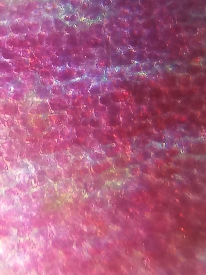
View in Media Gallery
Cherry peel – pigment inside the cells
View in Media Gallery
Cherry peel – pigment inside the cells
He shared the images on our local foldscope WhatsApp group.
Now starts the replication experiment and getting more clarity.
Mitali:
I was excited seeing the pictures sent by Deepak sir!
It was very intriguing the way the cells appeared packed and without the pigment !! Or pigment outside ?
I wanted to see it myself and verify it.
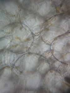
I rushed and stole a tomato from the kitchen and tried it out myself. I peeled the tomato skin and focused on the thinnest peel I could get.
The peel was just one cell layer thick! And I was taken for a shock. The cells had no pigement inside it at all.
He shared the images on our local foldscope WhatsApp group.
Now starts the replication experiment and getting more clarity.
Mitali:
I was excited seeing the pictures sent by Deepak sir!
It was very intriguing the way the cells appeared packed and without the pigment !! Or pigment outside ?
I wanted to see it myself and verify it.
I rushed and stole a tomato from the kitchen and tried it out myself. I peeled the tomato skin and focused on the thinnest peel I could get.
The peel was just one cell layer thick! And I was taken for a shock. The cells had no pigement inside it at all.
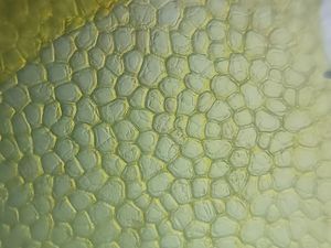
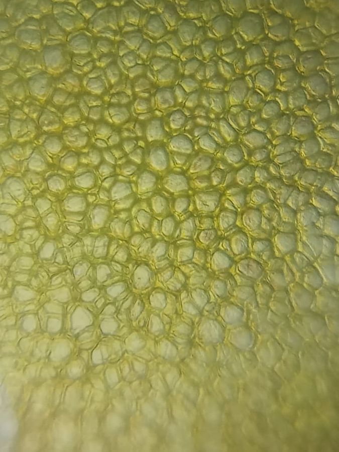
Tomato Peel (I see some large organelles in the cells but no pigment at all! Except for something at the periphery- I’m thinking it’s the Cell wall) This is how we landed up replicating the experiment and proved that tomato skin cells have no pigment
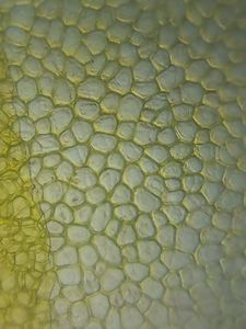
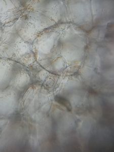
Focusing a thicker region made the skin appear yellow.
Focusing a thicker region made the skin appear yellow.
View in Media Gallery
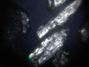
Thick tomato peel – for some reason I don’t see multiple cells stacked to give a mesh-like look Here is a picture of exactly 2 layers of cells.
View in Media Gallery
The peel that was single cell layer thick folded on itself looking very different from the thicker peel above) Now I was curious to figure out what exactly is making the tomatoes red?
Me and Deepak sir both individually took a look at the pulp
Me and Deepak sir both individually took a look at the pulp
View in Media Gallery
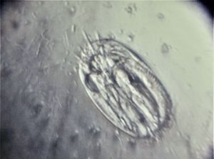
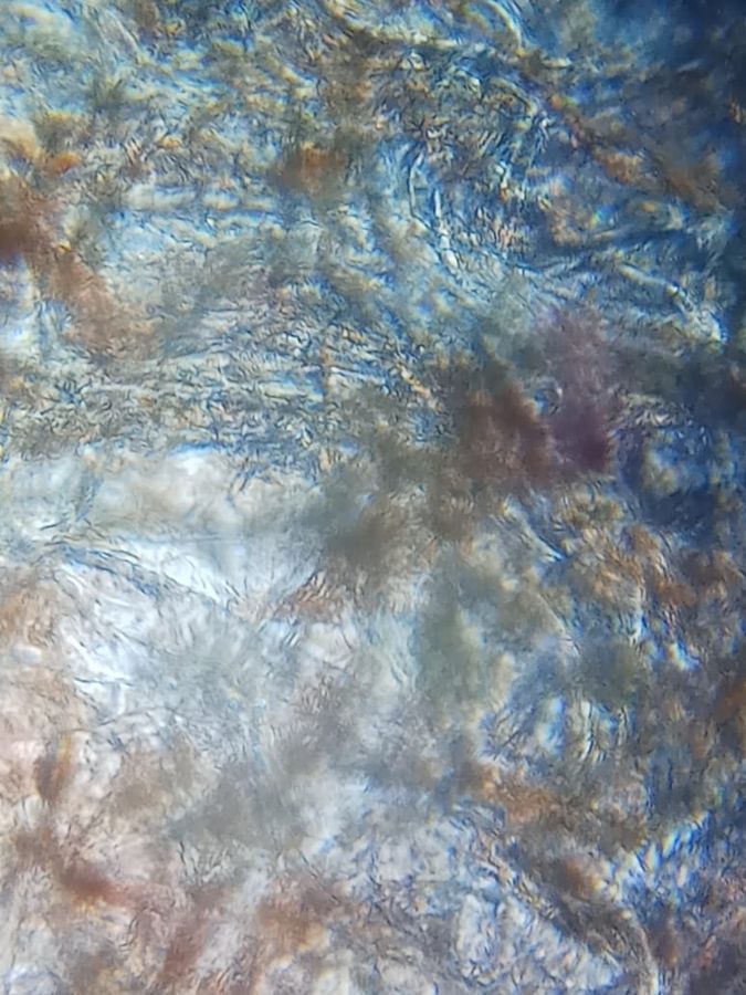
Scooping out the pulp was destroying the cells. He says they are fibers or fibrous cells. Nevermind. But I could still see some intact cells in the pulp so I decided to try sectioning the tomato.
And here are the results!
And here are the results!
You can see some organge/red granules in the cells at the periphery
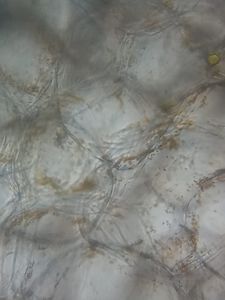
I got much better pictures of the cells and they were in great shape!
I could also see some granules inside these ‘pulp cells’ which I think are chromoplasts.
Chromoplasts are like chloroplasts except they store carotene, xanthophyll and lycopene pigements as opposed to chlorophyll in chloroplasts.
(I googled to find out which pigment is it exactly – It is Lycopene.)
So this is our story of how we found out – what gives tomato it’s red colour? 😀
Tomatoes have seprate chromsoplasts which gives its colour and not its skin cells.
And that this is not how cherries get their red colour.
Some tips for better imaging
– I used 3 layers of scotch tape on my light module to diffuse the light. (more on diffusing light by Manu sir here .)
– I mounted my sample in glycerol. It gives better images because of its refractive index.I always keep a bottle handy. It is cheap and one bottle lasts very long. However, I have never really compared the kind of data I get – Water vs Glycerol. (looks like a title for the next post!)
Thanks for reading!
– Deepak sir, Shradha and Mitali
Sign in to commentNobody has commented yet... Share your thoughts with the author and start the discussion!

 0 Applause
 0 Comments