Comparing stomata & epidermal cells between different species of plants
 Jul 09, 2019 • 8:13 PM UTC
 Unknown Location
 140x Magnification
 Microorganisms
Jay Holmes
I work at the American Museum of Natural History and love exploring the natural world at all scales.
2posts
86comments
1locations
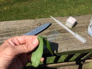
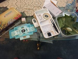
The lab: Foldscope kit and container of a variety of collected leaves. Collecting & Preparing a sample After reading the post by KAVITHA ( https://microcosmos.foldscope.com/?p=160084) about stomata, I had a question: Do all stomata look the same? So over the weekend, while visiting my sister and parents, we went on a Stomata Expedition! We collected a several different sorts of leaves, placing them in a plastic tub to keep them from drying out, and took them inside to examine them with the Foldscope!
The leaf is too thick and contains too many layers of cells to clearly see the stomata on the lower surface of the leaf, so we must peel the lower surface off of the leaf. We found the best way to do this was to fold the leaf so that the bottom surface of the leaf is on the outside of the fold.
The leaf is too thick and contains too many layers of cells to clearly see the stomata on the lower surface of the leaf, so we must peel the lower surface off of the leaf. We found the best way to do this was to fold the leaf so that the bottom surface of the leaf is on the outside of the fold.

View in Media Gallery
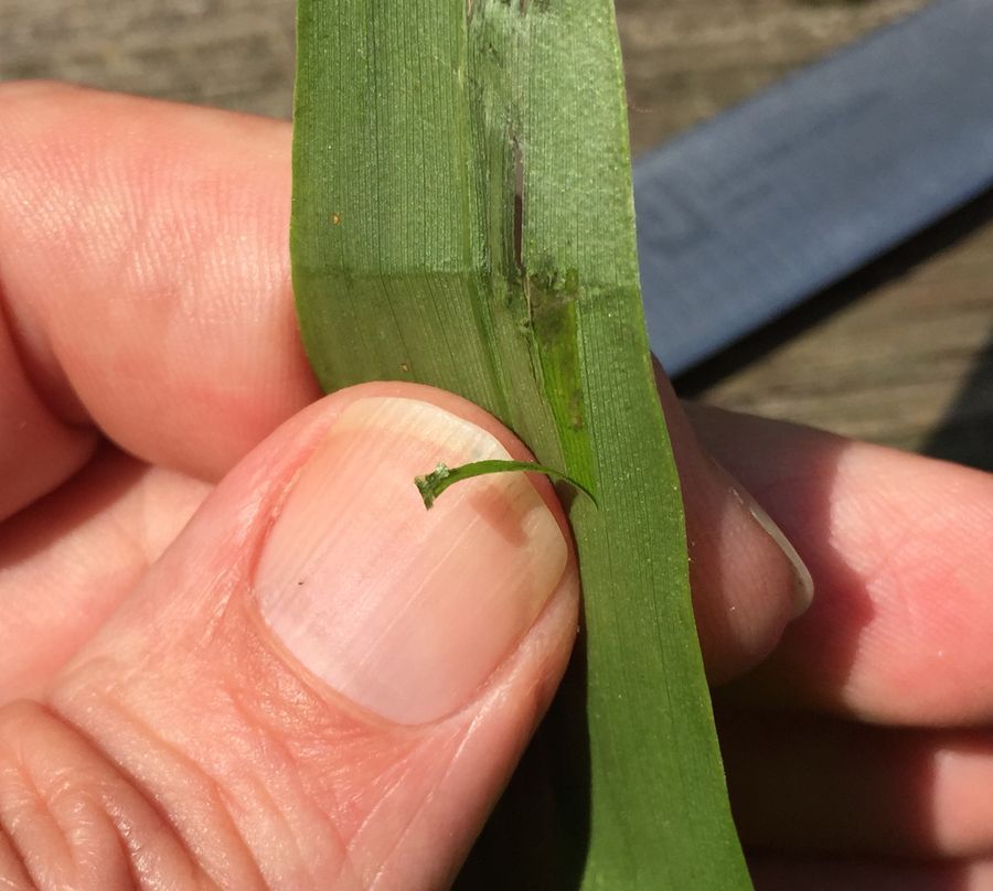
Folded leaf of Dichanthelium latifolium, Broad-leaved Panic Grass, bottom side out. The next step is to pinch the edge of the fold between your fingernails and try to peel the bottom layer of the leaf down, away from the fold.
View in Media Gallery
This pulled away little skin of the leaf still has some of the green inner cells (the spongy mesophyll) on it. We will scrape that off so we just have the clear epidermal cells, with the stomata, on the slide. I used my thumbnail to lightly scrape the green mesophyll off the inner side of the peel and then placed the thin, single cell thick, layer of leaf epidermis on a slide with a little water, then covered it with a cover slip.
View in Media Gallery
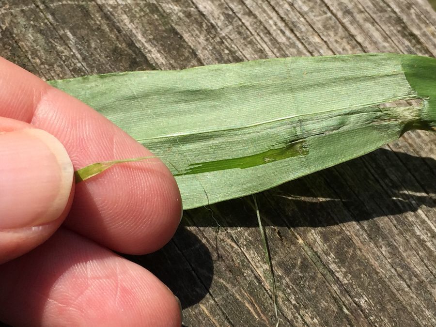
The thin scraped peel is slightly curled as it dries quickly in the air.
View in Media Gallery
Here you can faintly see the very clear peel under the coverslip. It is greener to the left were I didn’t get the green mesophyll peeled off. We had a variety of plants that we collected from the side of the road and around the house. And made peels of each. I will add picture of each plant in the field and what the stomata look like.
View in Media Gallery
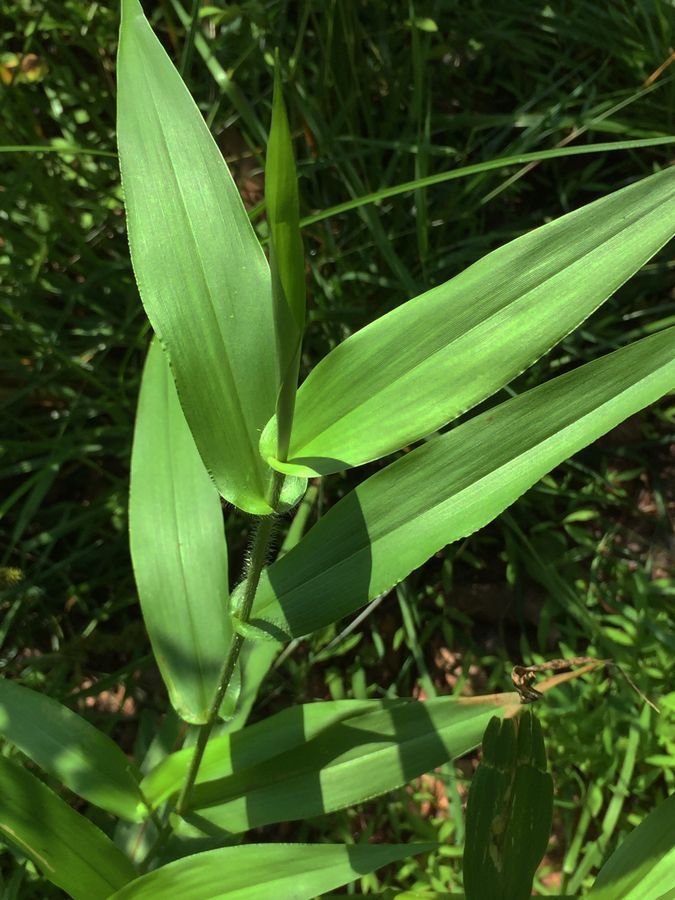
Dichanthelium latifolium, Broad-Leaved Panic Grass, a monocot.
View in Media Gallery
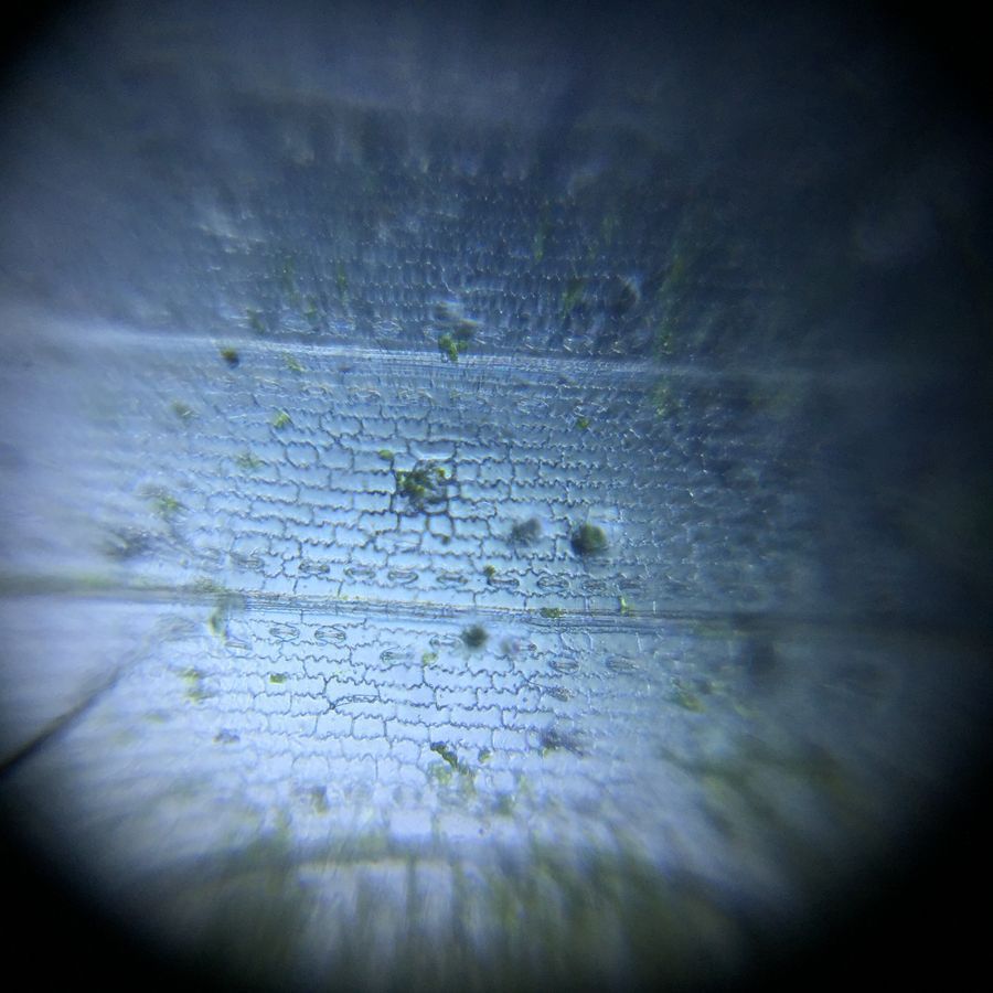
Dichanthelium latifolium, Broad-Leaved Panic Grass. In this one the stomata (the little oval things) appear in rows on either side of a rib in the leaf. Can you see them? there are 4 nicely visible rows in the middle 1/3 of the photo and others that are a little blurry towards the top and bottom.
View in Media Gallery
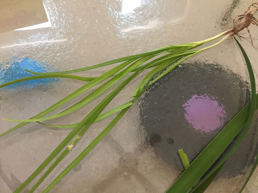
This is the Garlic Chive, Allium tuberosum , my sister pulled from her garden. I didn’t get a photo of it growing outside. It is another monocot.
View in Media Gallery
Epidermis of Allium tuberosum. The things that look like little mouths are the stomata. This image is zoomed in a little more on my phone, so they look a little bigger. How is this leaf similar to the first leaf? Can you see the small round dots inside some of the longer cells? These are cell nuclei. There are 3 or 4 nicely visible near the center of the image. Pretty cool!
View in Media Gallery
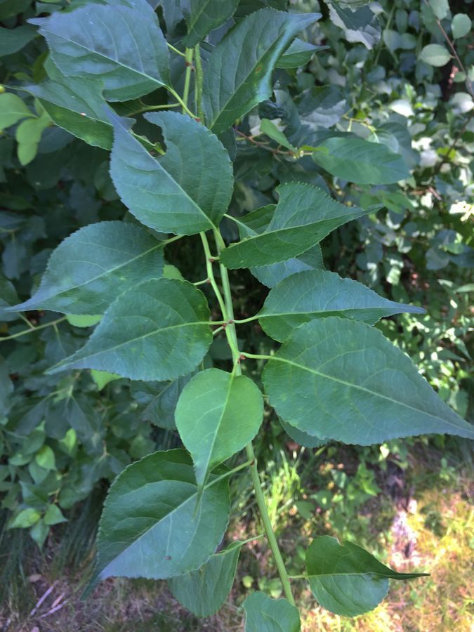
This one is Celastrus orbiculatus commonly known as Oriental Bittersweet around here. These leaves are broad, not long and slender like the previous two. This plant is in the eudicots, meaning it has a seed with two halves like a bean or a pea.
View in Media Gallery
What do you notice first about this leaf? The first thing that I noticed was NO ROWS! This one looks pretty messy! And the cells of the leaf are not generally rectangles and lined up like bricks, they are like puzzle pieces and appear fairly randomly arranged.
View in Media Gallery
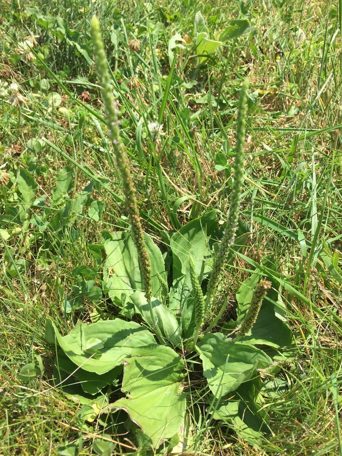
Plantago major , broadleaf plantain, another eudicot. What do you think the epidermal cells and stomata will look like?
View in Media Gallery
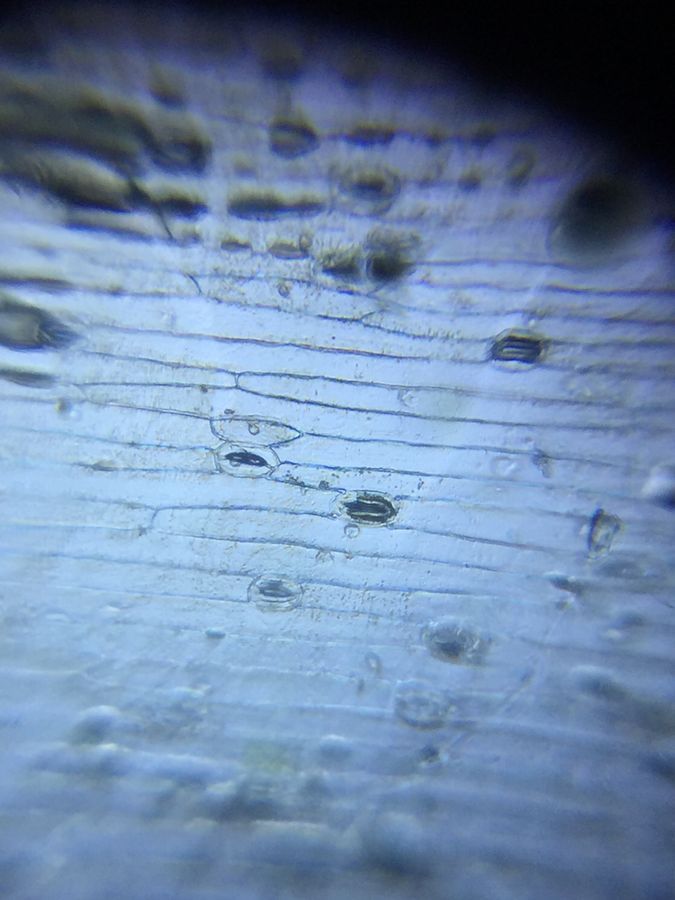
Plantago major, broadleaf plantain, epidermal cells and stomata. Which do these look most similar to, Celastrus orbiculatus or Allium tuberosum above?
View in Media Gallery
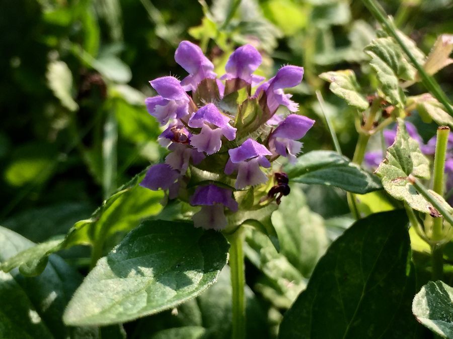
I like the look of this little flower that was growing in the yard. It is called Prunella vulgaris, or Common Selfheal. It is another eudicot, guess what?
View in Media Gallery
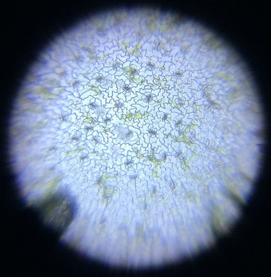
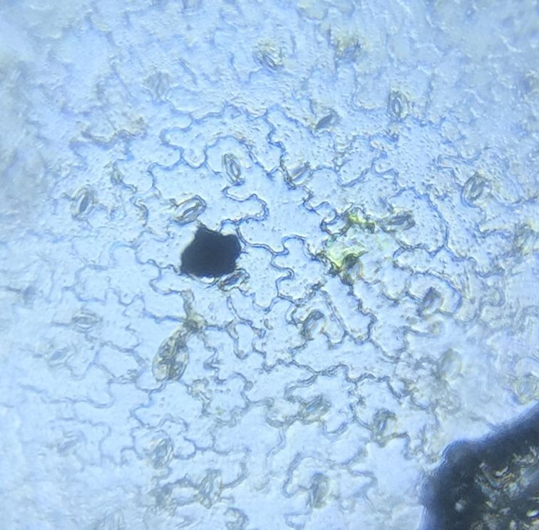
Prunella vulgaris has sort of random puzzle piece cells, like the other eudicots, but what do you notice about the stomata here? I think there are LOTS more on this leaf than the other eudicots. I wonder why? The green/dark blobs in here are some of the mesophyll cells that I did not remove.
View in Media Gallery
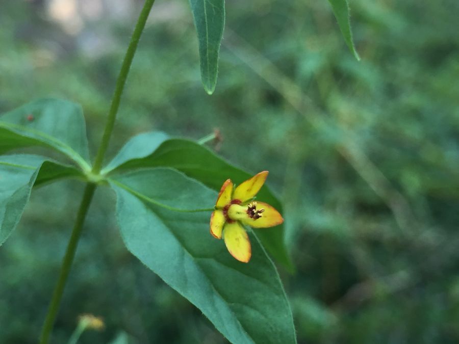
Last plant! This one is Lysimachia quadrifolia , the Whorled Loosestrife. Look at the leaf, which group do you think that it might be in? What do you think the epidermis might look like?
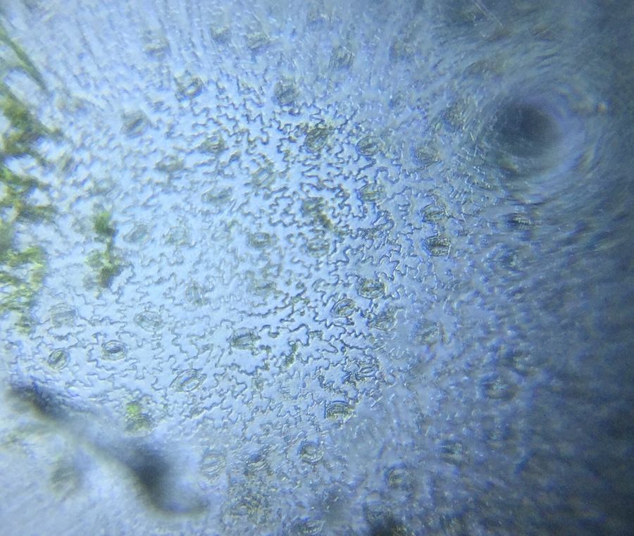
View in Media Gallery
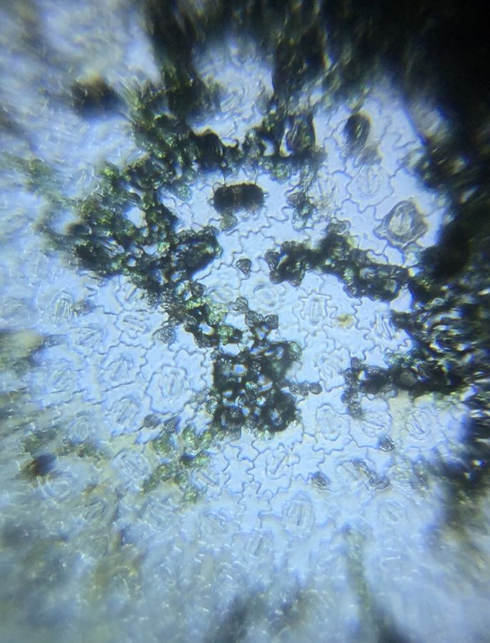
The Epidermis of the Lysimachia quadrifolia , Whorled Loosestrife. With the puzzle piece shaped cells and the scattered stomata, similar to the other eudicots. So the answer to my question was, at this magnification, the stomata look fairly similar between the plants, but they are arranged very differently in different larger groups of plants and between species within these large groups of plants. There seemed to be some leaves with MANY stomata and some with not so many. And the shapes and arrangement of epidermal cells seem to be particular to whether the plant was a monocot or a eudicot. What questions do you have about these observation? What would you look at next?
I found this great fun. A few things that I learned were, scraping the mesophyll cells off helps, they are thick and can make focusing tricky. I also learned that the focus can shift a bit when I go from viewing with my eye to taking a picture with the phone, so that takes a few tries, and toss out the poorly focus images. Cropping out the out-of-focus parts around the edge is helpful, but it is hard to get a sense of how big things are relative to each other if different images are cropped differently.
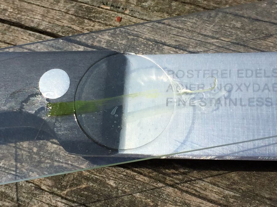
For this project I was using glass slides and coverslips, the Foldscope LED illuminator directed straight through the lens, and an iPhone 6, mostly at full frame for photographing, then cropped. I used a knife to trim the leaf material so the piece that was left was just a thin piece of epidermis.
I found this great fun. A few things that I learned were, scraping the mesophyll cells off helps, they are thick and can make focusing tricky. I also learned that the focus can shift a bit when I go from viewing with my eye to taking a picture with the phone, so that takes a few tries, and toss out the poorly focus images. Cropping out the out-of-focus parts around the edge is helpful, but it is hard to get a sense of how big things are relative to each other if different images are cropped differently.
For this project I was using glass slides and coverslips, the Foldscope LED illuminator directed straight through the lens, and an iPhone 6, mostly at full frame for photographing, then cropped. I used a knife to trim the leaf material so the piece that was left was just a thin piece of epidermis.
Sign in to commentNobody has commented yet... Share your thoughts with the author and start the discussion!

 0 Applause
 0 Comments