My housemate’s Pancake just shed…
 May 16, 2016 • 11:36 AM UTC
 Unknown Location
 140x Magnification
 Microorganisms
Clifton Herrmann
Learn about the author...
4posts
0comments
2locations
View in Media Gallery
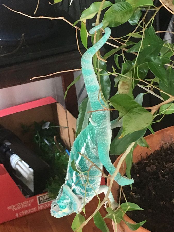
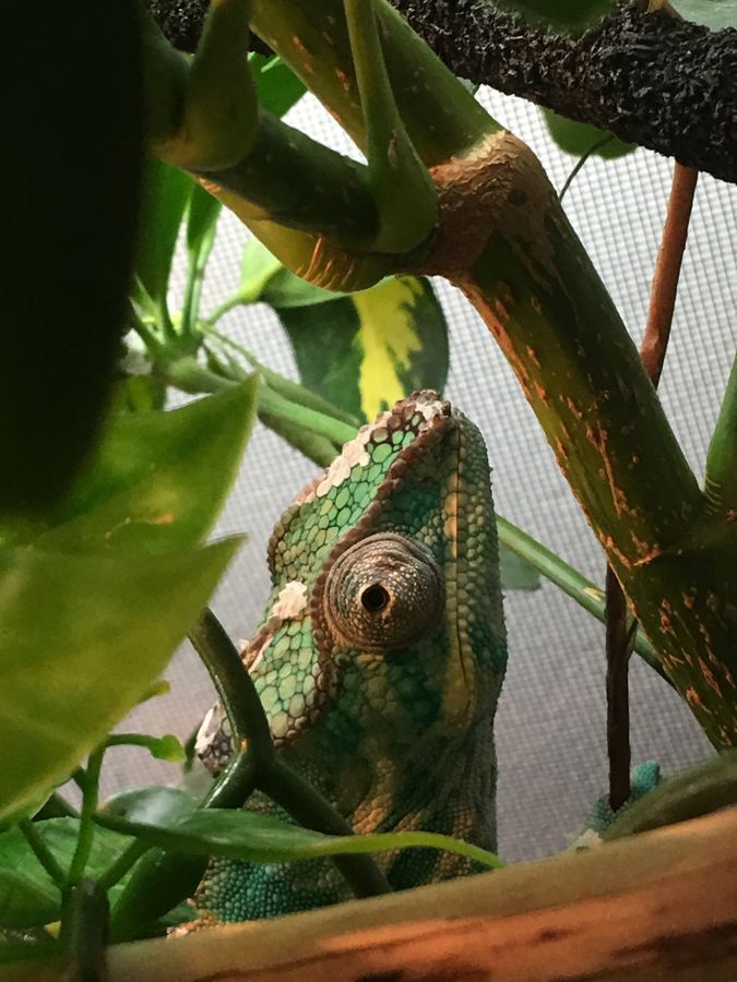
In past housing situations I’ve been lucky to enjoy play-only relationships with dogs whose physical and health needs were otherwise met by their responsibility-laden owners. Having recently moved into a new shared rental, I’m experiencing a unique treat in that one of my housemates is a true-story Chameleon owner. Pancake is a stunning-in-teal Panther Chameleon, species Furcifer pardalis . This species originates in northern Madagascar and is prized by exotic pet owners for its bright display of color-changing skin.
View in Media Gallery
As an ecologist I have enjoyed watching Pancake transform from teal-pink-yellow to marble white as he falls asleep in the evening. And as a marine ecologist, I’ve found myself assuming the mechanism behind that change to be something familiar to me; organisms such as the octopus and squid that change color by stretching or contracting pigment-filled cells can be a common topic of interest. The more I thought about this, however, the more I slowly started to realize that Pancake’s skin is drastically different than those miraculous mollusks..
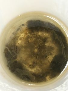
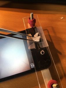
Luckily this week he decided to shed some healthy looking skin, and I went straight to work on my FoldScope with a fresh piece!
Luckily this week he decided to shed some healthy looking skin, and I went straight to work on my FoldScope with a fresh piece!

View in Media Gallery
I set up my new popsicle stick FoldScope rig, fired up the flashlight

View in Media Gallery
And just like that I was getting some great shots!
View in Media Gallery
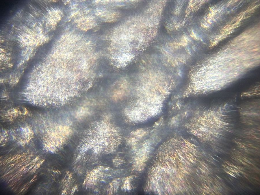
I found that by keeping the sample still and rotating the flashlight so that the direction and distance of the light source shifted, the skin would refract a beautiful kaleidoscope of colors! Right away I could tell I was onto something with my color-changing question. If pigment-based chromatophores were causing the color change, why would the skin left behind be so dang colorful? There had to be more to it…

View in Media Gallery
In my research I found that it really was refraction rather than some sort of actual pigment! Turns out they have five main types of chromatophores: some are pigment based and occur in the deeper epidermis (Carlton 1903), and others actually contain microscopic crystals that disperse/aggregate based on the tightness of its skin (similar to the octopus, but using crystals instead of pigment!). These guanine nanocrystals are covered by a thick superficial layer of dermal iridophores, resulting in two interactive layers of refractive material (Teyssier et al, 2015)!
Holy Chameleon! How awesome! Turns out Pancake is actively utilizing the physical phenomenon of refraction to dictate his color displays, ultimately describing his emotions to me! Talk about wearing your heart on your sleeve….
Holy Chameleon! How awesome! Turns out Pancake is actively utilizing the physical phenomenon of refraction to dictate his color displays, ultimately describing his emotions to me! Talk about wearing your heart on your sleeve….
View in Media Gallery
References
Carlton F.C. 1903. The color changes in the skin of the so-called Florida Chameleon, Anolis carolinensis . Proceedings of the American Academy of Arts and Sciences. 39(10):259 – 276.
Teyssier J., S.V. Saenko, D. Van Der Marel, M.C. Milnikovich. 2015. Photonic crystals cause active colour change in chameleons. Nature Communications. DOI: 10.1038/ncomms7368.
Sign in to commentNobody has commented yet... Share your thoughts with the author and start the discussion!
 0 Applause
 0 Comments