Microbial diversity in a paddy field water sample – Part 3
 Aug 05, 2019 • 1:53 AM UTC
 Unknown Location
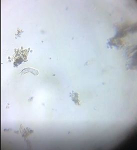
 140x Magnification
 Microorganisms
Jayashree Ramadas
We are a group of students, volunteers and staff working with TIFR Hyderabad's Science Education and Outreach program: http://www.tifrh.res.in/~outreach/
39posts
26comments
2locations
View in Media Gallery
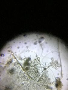
TWO WORMS
View in Media Gallery
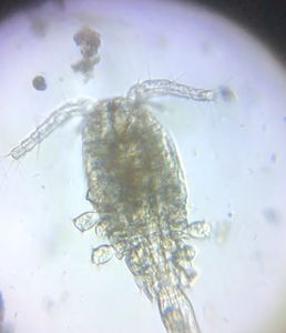
Nematodes were in the sample right from Day 1. I was a bit neglectful of them perhaps because, in the numerous samples that I have observed till today, this guest has always been ready to come under my Foldscope. It was on Day 2 that I observed two new worms , one with long bristles on either side of the body, while the other one was busy in grabbing food.
View in Media Gallery
At first, I thought the one with bristles was an Annelid worm and the other busy one might be a rotifer (its feeding behaviour looks like that)!! However, I could not find the mastax organ in its pharynx. The lower worm was smaller, and it did not have segments or setae. But leaving it aside for now, I tried to focus on the Annelid worm (with bristles) and closely scanned its body through a Foldscope video.
Except for the head, the other segments had a set of long bristles (setae) and the Prostorium (head portion) was broader than the body. I tried to count the segments – there were around seventeen. Whenever I shone the light source under the head, it turned away or buried its head under the algal filaments. This worm was definitely sensitive to light.
I had studied peristalsis only through words in the textbook. It was an amazing experience to watch the peristaltic movement in the dorsal and ventral blood vessels, and in the digestive system of this worm. There were some orange-coloured droplets in its coelom. From these observations, I came to the conclusion, that it is probably an Aeolosoma sp .
View in Media Gallery
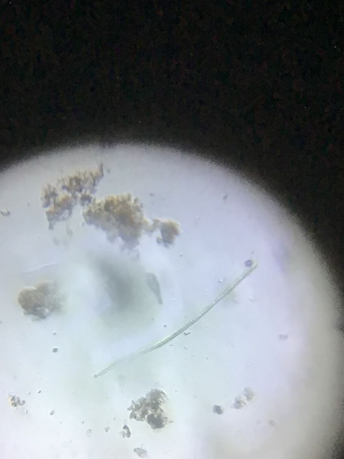
Unfortunately, while observing the first one, I missed the second worm. I tried for a few days to find it but with no success. After a few days, suddenly I got one worm-like movement in between the debris. Was it a baby Aeolosoma? Probably not! There were no long bristles or segmentation in its body. I could observe clearly the muscle contraction and relaxation, while it was feeding and the muscles were forcing down the food towards the posterior end through a peristaltic movement. I think it is probably Stenostomum sp., a flatworm belonging to Turbellaria. This video on YouTube looks similar.
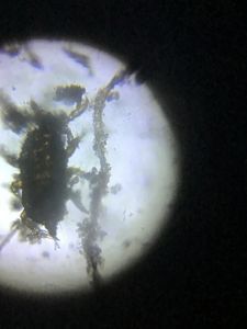
ARTHROPODA
ARTHROPODA
View in Media Gallery
All through the days of observation, I found arthropods.
View in Media Gallery
I located a Nauplius (larval stage of a Copepod) which did a sudden vanishing act (at 00:24 sec).
View in Media Gallery
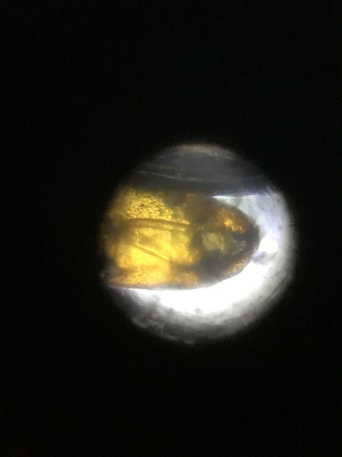
There was a yellow coloured arthropod with big eyes, but I was unable to locate any antenna, to confirm it as a copepod. Is it a Copepod?
View in Media Gallery
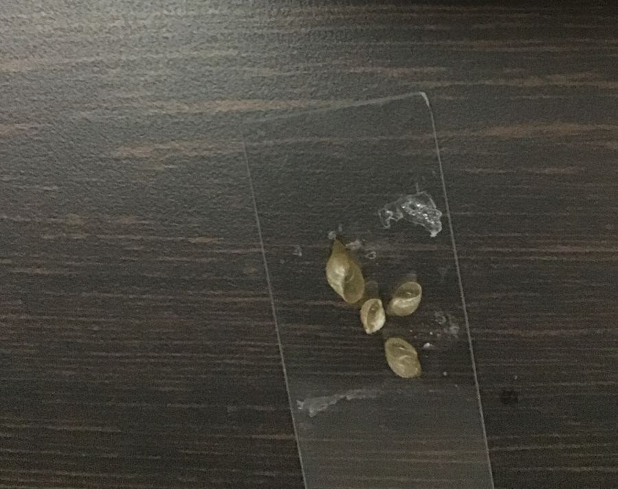
MOLLUSCA Under the magnifier lens I saw some empty dextral gastropod shells.
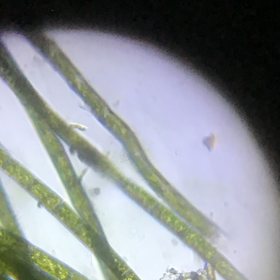
PHYTOPLANKTON Well! So far I have discussed my observations about zooplanktons only, which included different levels of consumers. But what about the producers? While observing these moving microbial organisms, I did notice a number of wonderful phytoplanktons as well. These producers belonged to both prokaryotes and eukaryotes.
PHYTOPLANKTON Well! So far I have discussed my observations about zooplanktons only, which included different levels of consumers. But what about the producers? While observing these moving microbial organisms, I did notice a number of wonderful phytoplanktons as well. These producers belonged to both prokaryotes and eukaryotes.
View in Media Gallery
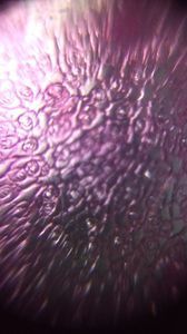
a) I observed a type of Cyanobacteria , with four bluish-green hemispherical cells, enclosed within a mucilage sheath. I s it a Chroococcus ?
View in Media Gallery
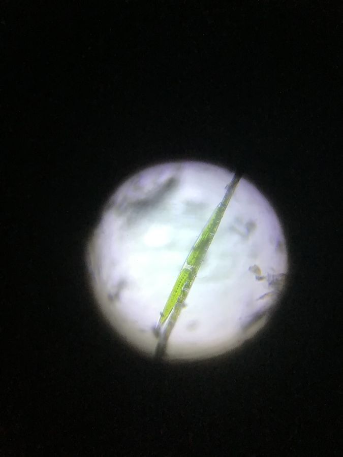
There was also one more distinctive Cyanobacteria, i.e Oscillatoria sp . It is a long filamentous bright bluish-green cyanobacteria, resembling a green snake, which shows oscillating and gliding movements.
View in Media Gallery
Through its slight oscillatory movement , I concluded its identity as an Oscillatoria sp .
View in Media Gallery
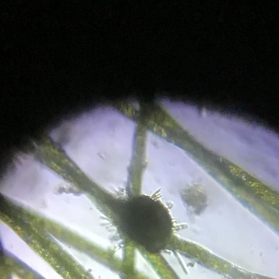
b) I also observed diatoms , which are eukaryotes usually found in water samples. They were bright yellowish-brown and unicellular, probably pennate diatoms. Usually diatoms are non-motile, however, I noticed that these diatoms were moving, or perhaps just gliding due to the water movement.
View in Media Gallery
c) Another interesting observation was a Closterium , which is a unicellular organisms with tapering ends at both sides, consisted of two chloroplasts and plenty of pyrenoids (a central line of dark green spots). These are the characteristics of
desmid Closterium.
desmid Closterium.
View in Media Gallery
d) I also observed Euglena , a eukaryote with flagella and chloroplasts. From its elongated shape, tapering end, red eyespot and characteristic movement (as I found in other videos) I identified it as Euglena.
View in Media Gallery
Movement of euglena
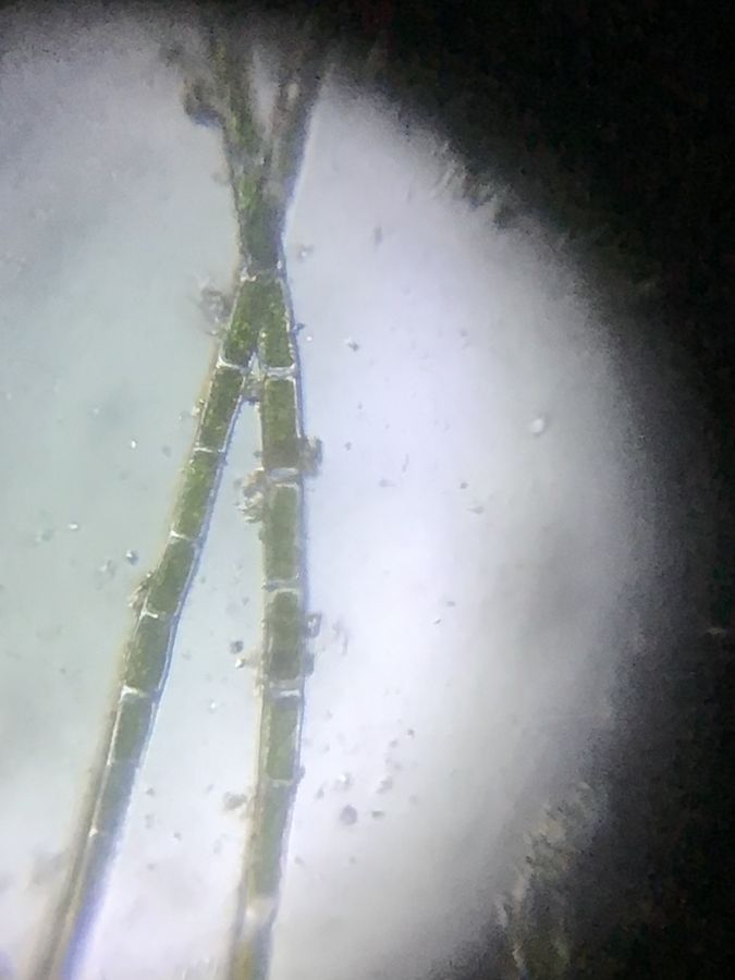
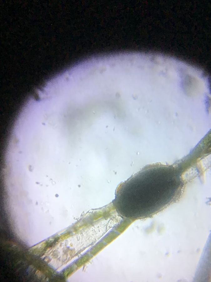
Finally, I observed some unbranched Oedogonium filaments with cap cells, having reticulate chloroplasts which fully occupied the cells (see Image 1). I observed a beautiful diatoms colony on the filament (see Image 2). I could confirm that the Oedogonium sp. is nannandrous , from the few-celled Antheridial (male) dwarf filaments attached to the main filament. I assume that this species is idioandrosporous because I couldn’t find any oogonium near the Antheridial dwarf filament (see Image 3). I also observed the oogonium with zygote surrounded by a thick wall (see Image 4).
Finally, I observed some unbranched Oedogonium filaments with cap cells, having reticulate chloroplasts which fully occupied the cells (see Image 1). I observed a beautiful diatoms colony on the filament (see Image 2). I could confirm that the Oedogonium sp. is nannandrous , from the few-celled Antheridial (male) dwarf filaments attached to the main filament. I assume that this species is idioandrosporous because I couldn’t find any oogonium near the Antheridial dwarf filament (see Image 3). I also observed the oogonium with zygote surrounded by a thick wall (see Image 4).
View in Media Gallery
Image 1 – Oedogonium filament with capcells
View in Media Gallery
Image 2 – Diatoms colony on Oedogonium filament
View in Media Gallery
Image 3 – Dwarf male filament of Oedogonium filament
View in Media Gallery
Image 4 – Oogonium with zygote I had a wonderful experience with this water sample with wide biodiversity (see also Part 1 and Part 2 of the series). Thank you to the foldscope team which gave me an opportunity to observe and to take videos. These videos gave me time to observe freely and helped me to improve my knowledge.
Cheers!
Ashalatha
with Debashree, Jayashree, Chandrika
Cheers!
Ashalatha
with Debashree, Jayashree, Chandrika
Sign in to commentNobody has commented yet... Share your thoughts with the author and start the discussion!

 0 Applause
 0 Comments