Skeleton Shrimp (Caprella) – day 2 of Hopkins course
 May 26, 2016 • 10:45 PM UTC
 Unknown Location
 140x Magnification
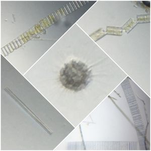
 Microorganisms
Saad Bhamla
Learn about the author...
32posts
11comments
2locations

View in Media Gallery
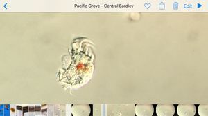
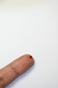
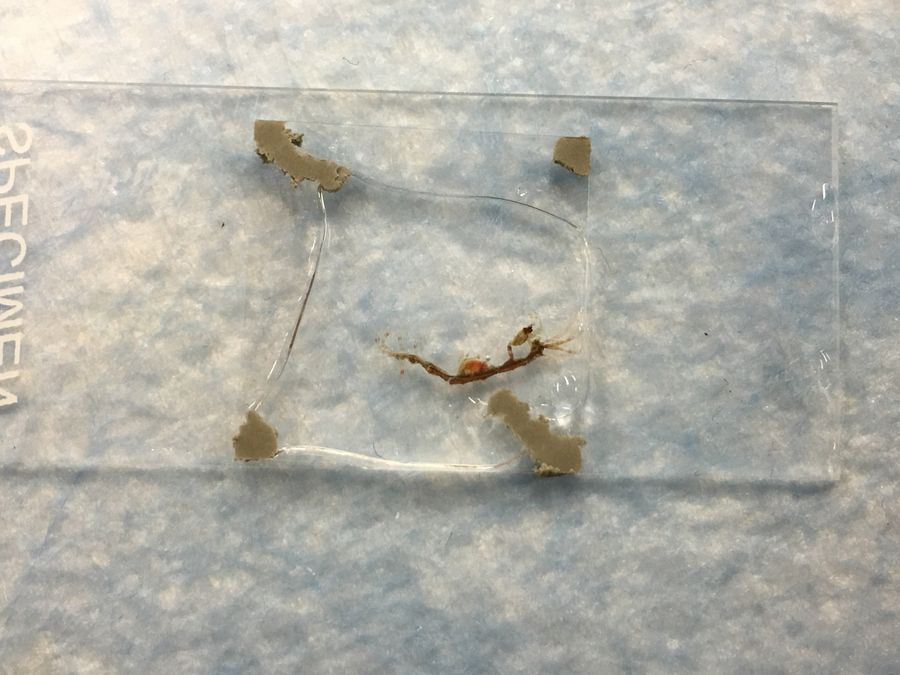
Today i managed to put it in a skeleton shrimp in my foldscope.
View in Media Gallery
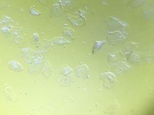
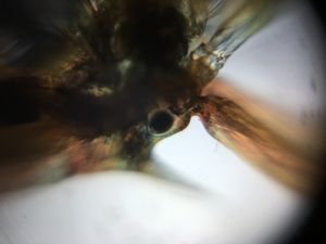
Here’s a video showing blood flowing through the shrimp. It’s incredible. You can see individual hemocytes speeding along.
I took a longer video with H, who studies these Caprella as a model organism, and she explained different features to us, including spotting a beating heart! I have to share a disclaimer that this particular shrimp is a female and we actually anesthetized, to extract the embryos, but looked like it wasn’t going to make it, and you’ll notice its heart beat is extremely low – i counted 2 beats separated by 8 seconds or so..
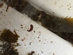
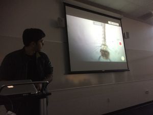
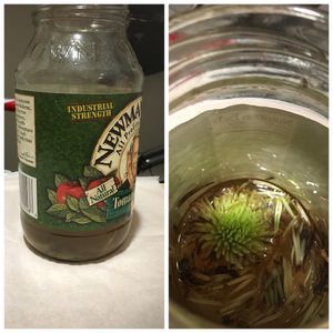
For those interested, here is a whole dissection video, but this was on a stereo microscope showing how we extracted the embryos for staining.
Finally, the class group is posting more pictures/videos live on to a class tumblr right here, in case you want to follow. I will share my foldscope videos and posts there, while simultaneously post long posts here directly on microcosmos too.
Link: http://hopkinsembryology2016.tumblr.com
Link: http://hopkinsembryology2016.tumblr.com
Sign in to commentNobody has commented yet... Share your thoughts with the author and start the discussion!

 0 Applause
 0 Comments