How can jellyfishes sting?
 Aug 08, 2019 • 2:56 PM UTC
 Unknown Location
 140x Magnification
 Microorganisms
Cristina Bosch Esteva
Learn about the author...
19posts
52comments
1locations

View in Media Gallery
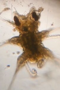
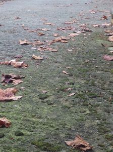
My city has a wonderful nearby beach known as Las Canteras beach. Last wednesday there were many jellyfishes on the sea shore belonging to Pelagia noctiluca species, cnidarians that the waves drag and leave on the sand when they bloom due to higher sea temperatures, among other reasons.
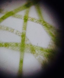
I have always wondered how cnidocites (stinging cells that these animals have) look like under a microscope. Therefore, I carefully picked two of them and took them home.
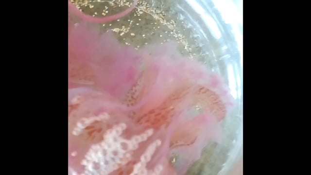

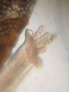
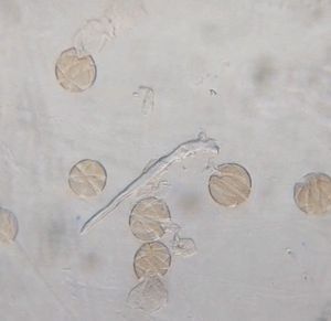
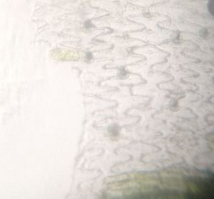
I viewed a tentacle (pointed above) and saw clusters and clusters of spherical structures that represent cnidocites batteries! Watch by your own:
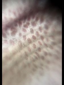

To my astonishment, many of these cells were detached from the body surface. I wonder if they can function under this circumstance.
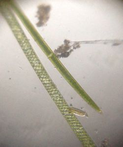
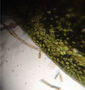
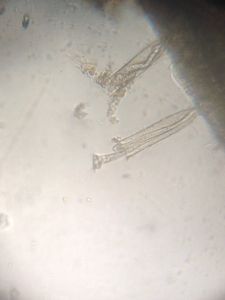
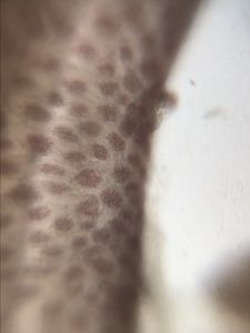
I could not see what these cells fire to act as stinging elements and was about to abandon when, wow!, I watched this:
I could not see what these cells fire to act as stinging elements and was about to abandon when, wow!, I watched this:
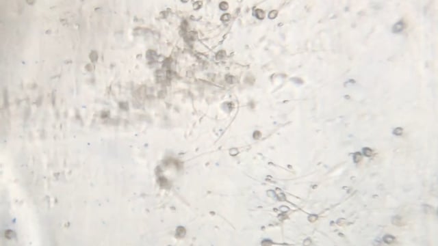

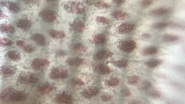
I can understand now that thousands of microscopic darts can sting in our skin all in a sudden carrying irritating or venomous substances. Another “discovery” achieved thanks to this incredible paper microscope!
Cheers!
Cristina Bosch
Cheers!
Cristina Bosch
Sign in to commentNobody has commented yet... Share your thoughts with the author and start the discussion!

 0 Applause
 0 Comments