A Tale of Two Ferns
 Jun 05, 2016 • 2:29 AM UTC
 Unknown Location
 140x Magnification
 Microorganisms
Max Coyle
Learn about the author...
16posts
17comments
3locations

View in Media Gallery
I have been playing with my foldscope for just a few days but have already found so much that is fascinating. As a molecular biology researcher and student, it has been refreshing to shift my attention from designing the perfect, incisive experiment to simply being curious about the miniscule details within the things all around me.
Representing this shift towards the experiential, it made sense to make my first Foldscope post about ferns: wondrous green comforting things that I know almost nothing about scientifically but which have always been my favorite group of plants.
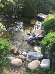
I first loved ferns growing up in the Pacific Northwest, and upon a brief return to Seattle (to meet up with my family en route to South Africa), I took the opportunity to collect samples from two ferns growing outside of the house I grew up in.
On the long transcontinental flight to South Africa, I investigated these ferns under the Foldscope. That I was able to collect interesting information using the Foldscope, while cramped in a small airplane seat, fumbling around on a tiny tray table, the earth wobbling below me, is a testament to the versatility and ease of the instrument.
The two ferns I explored are shown below, after mounting on the Foldscope paper slide. I have only used these paper slides so far, and have yet to compare to results with glass. I did use FOV locking and focus locking, with the LED light source and my iPhone camera.
Representing this shift towards the experiential, it made sense to make my first Foldscope post about ferns: wondrous green comforting things that I know almost nothing about scientifically but which have always been my favorite group of plants.
I first loved ferns growing up in the Pacific Northwest, and upon a brief return to Seattle (to meet up with my family en route to South Africa), I took the opportunity to collect samples from two ferns growing outside of the house I grew up in.
On the long transcontinental flight to South Africa, I investigated these ferns under the Foldscope. That I was able to collect interesting information using the Foldscope, while cramped in a small airplane seat, fumbling around on a tiny tray table, the earth wobbling below me, is a testament to the versatility and ease of the instrument.
The two ferns I explored are shown below, after mounting on the Foldscope paper slide. I have only used these paper slides so far, and have yet to compare to results with glass. I did use FOV locking and focus locking, with the LED light source and my iPhone camera.

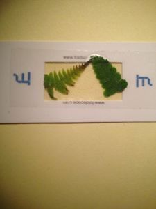
Fern A has that beautiful, classic taper so characteristic of ferns, and wider spacing between the leaves branching from the main stalk, compared to Fern B, which is lusher and more rounded.
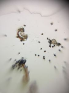
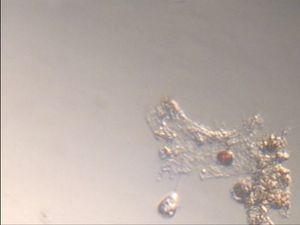
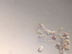
I have spent the last few years in lab studying the self-organization of microvasculature, and so naturally I was drawn to the vasculature of these ferns.
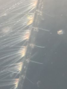
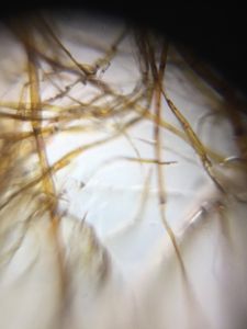
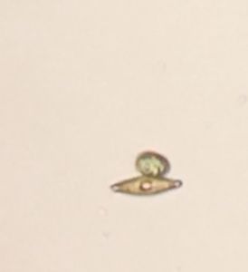
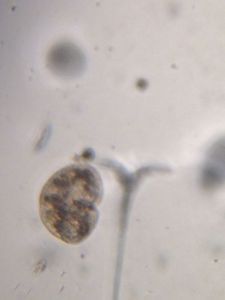
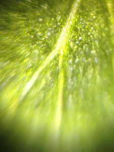
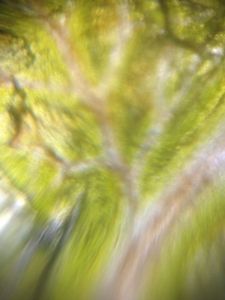
Fern B had a lovely, quite regular branching pattern to its vasculature. Each branch emerged from the pattern at the same angle every time, as shown in the pictures and video below.
I have spent the last few years in lab studying the self-organization of microvasculature, and so naturally I was drawn to the vasculature of these ferns.
Fern B had a lovely, quite regular branching pattern to its vasculature. Each branch emerged from the pattern at the same angle every time, as shown in the pictures and video below.
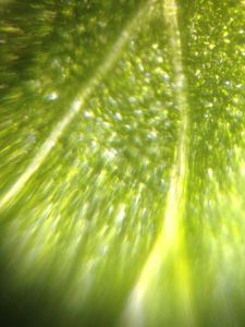
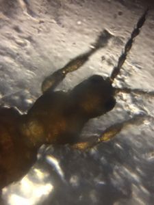
In contrast, the vasculature of Fern A branched more spasmodically and frequently, with many sharper turns involved.
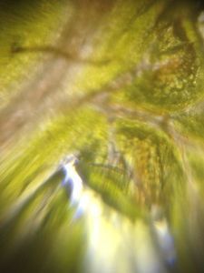
The difference was reminiscent of the difference between normal vasculature and tumor vasculature in animals, the latter more tortuous and ad hoc.
I know little about plant development and don’t have sophisticated theories about why these two ferns have different vasculature branching patterns. But I am curious to investigate more in the field and perhaps read up on vasculature in plants.
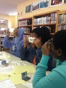
(I also brought some Foldscopes with me to South Africa and had the chance to put on a workshop for high school students, so consider this a teaser for my next post… )
I know little about plant development and don’t have sophisticated theories about why these two ferns have different vasculature branching patterns. But I am curious to investigate more in the field and perhaps read up on vasculature in plants.
(I also brought some Foldscopes with me to South Africa and had the chance to put on a workshop for high school students, so consider this a teaser for my next post… )
Sign in to commentNobody has commented yet... Share your thoughts with the author and start the discussion!
 0 Applause
 0 Comments