Mysteries of Dal Lake, Kashmir – Part 1
 Oct 16, 2019 • 6:51 AM UTC
 Unknown Location
 140x Magnification
 Microorganisms
Jayashree Ramadas
We are a group of students, volunteers and staff working with TIFR Hyderabad's Science Education and Outreach program: http://www.tifrh.res.in/~outreach/
39posts
26comments
2locations
View in Media Gallery
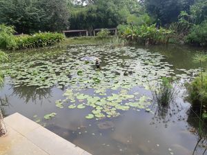
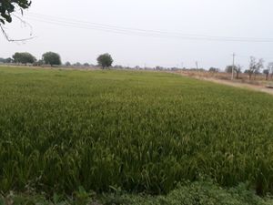
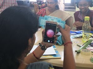
It all started when my friend got a water sample from Dal Lake, Srinagar. The ecosystem of Dal is ecologically rich with 117 recorded species of hydrophytes and phytoplankton . Here are some views of this beautiful Lake: wonder what we can find inside it, under the Foldscope!
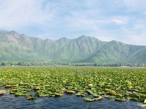
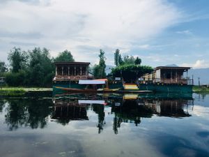
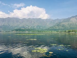
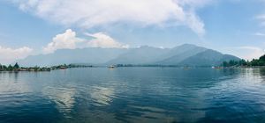
Dal Lake, Srinagar, Kashmir
View in Media Gallery
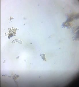
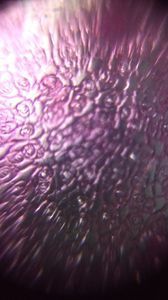
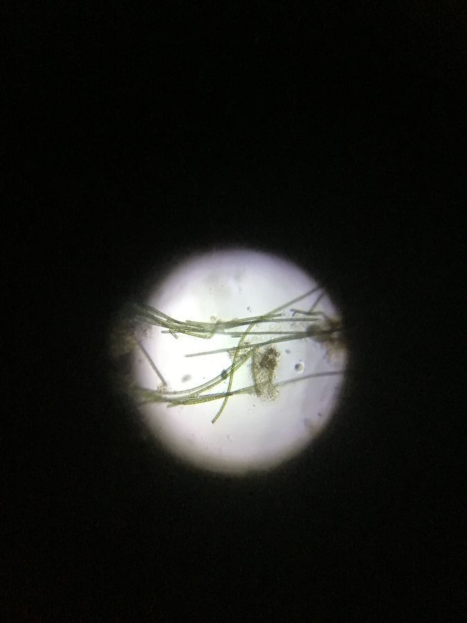
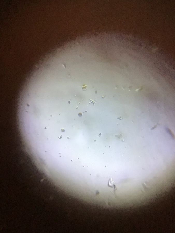
In my first observation, I saw many microbes moving in so many different ways! I used maximum zoom on the iPad to view them under the Foldscope.
View in Media Gallery
Look carefully at the centre of the field of view – the spiral motion is enthralling! This is perhaps a spiral bacterium. Isn’t it?
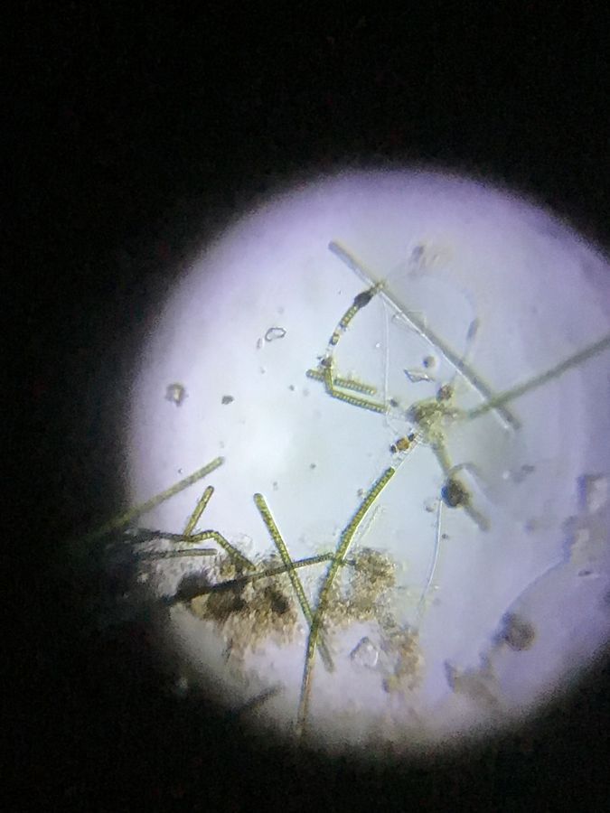
I found some prokaryotic filamentous cyanobacteria; they are bluish-green. In the second photo we can see an akinete on one of the filaments.
I found some prokaryotic filamentous cyanobacteria; they are bluish-green. In the second photo we can see an akinete on one of the filaments.
View in Media Gallery
Cyanobacteria
View in Media Gallery
Cyanobacteria
View in Media Gallery
Another type of cyanobacteria, Oscillatoria sp. , are capable of a wave-like motion. Notice the filaments oscillating back and forth.
We can see ‘separation discs’ (colourless or whitish) in the filaments.
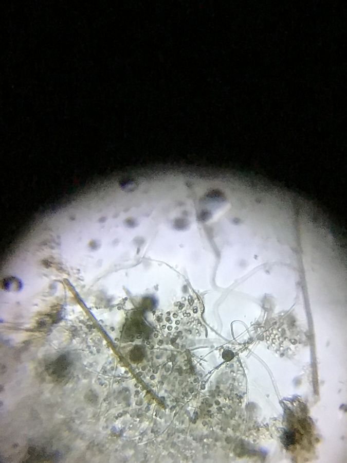
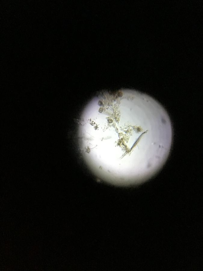
There was a variety of prokaryotic and eukaryotic life in this sample and a treat to see under the Foldscope! Can you help me identify some?
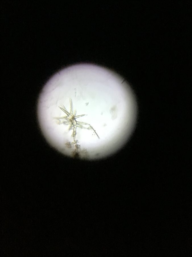
I think the star shaped structure is a diatom called Actinastrum and the cylindrical-stacked structure is a desmid called Scenedesmus .
We can see ‘separation discs’ (colourless or whitish) in the filaments.
There was a variety of prokaryotic and eukaryotic life in this sample and a treat to see under the Foldscope! Can you help me identify some?
I think the star shaped structure is a diatom called Actinastrum and the cylindrical-stacked structure is a desmid called Scenedesmus .
View in Media Gallery
Observe the star-shaped and cylindrical-stacked green structures.
View in Media Gallery
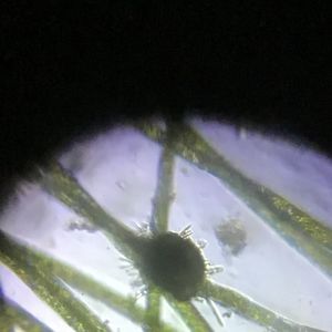
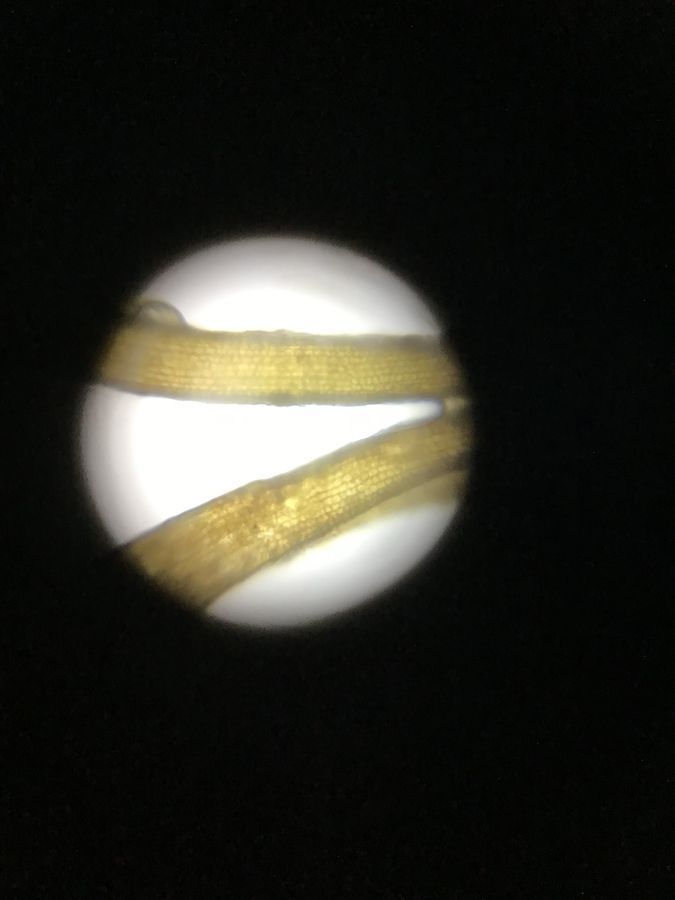
This rib-like structure is a perhaps a diatom too.
View in Media Gallery
Is this a diatom?

View in Media Gallery
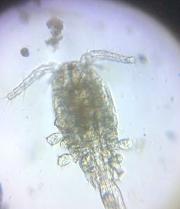
From these spiral choloroplasts I deduced this one is Spirogyra sp . A dead rotifer seems to be floating around.
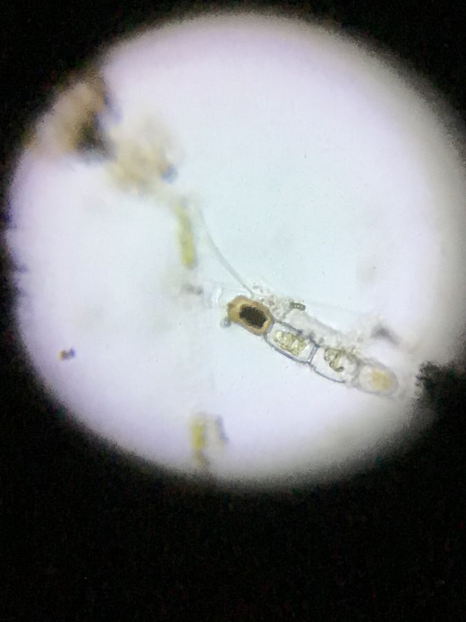
View in Media Gallery
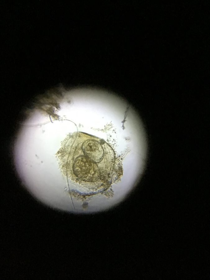
I was thrilled to see the ladder-like structure (one filament seems empty and the other is with a zygote). I believe this is the scalariform conjugation of Spirogyra . Can anyone help to verify this?
View in Media Gallery
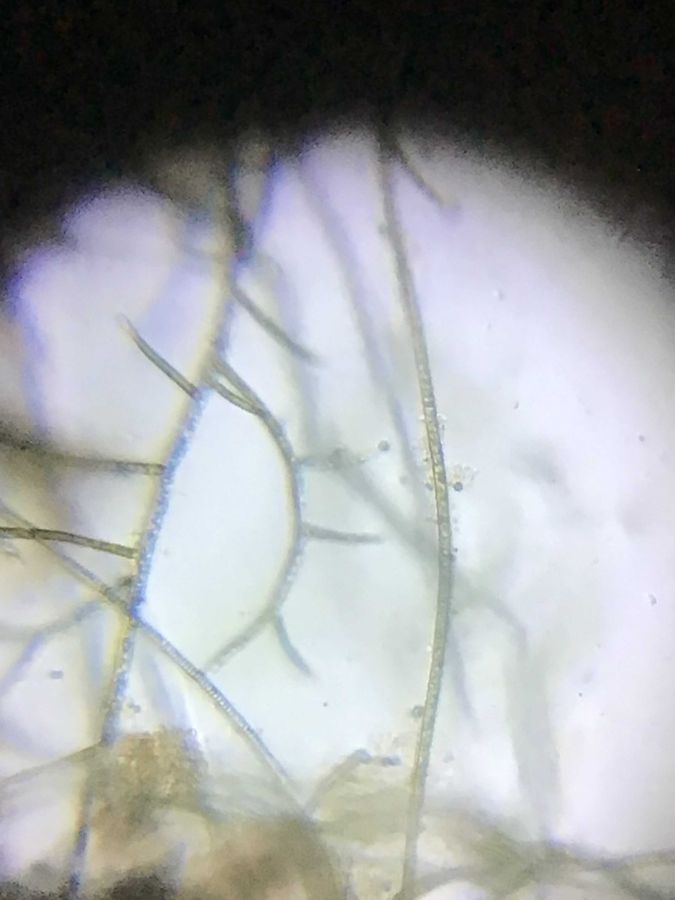
I also saw branched algae. I think this is Stigeoclonium sp.. Is it correct?
I found plant-like algae in the water sample. The image on the left is observed under the magnifying glass attached to the Foldscope light source. I think it is Chara sp .. The picture to the right is the detail of the leaf-like structure under the Foldscope. (I use concave slides to examine water samples!)
I found plant-like algae in the water sample. The image on the left is observed under the magnifying glass attached to the Foldscope light source. I think it is Chara sp .. The picture to the right is the detail of the leaf-like structure under the Foldscope. (I use concave slides to examine water samples!)

View in Media Gallery
Plant-like algae – Could it be Chara ?
View in Media Gallery
This is the leaf-like structure There were many structures or algae that I could not distinguish. They are so unique, different from any that I have seen before. What do you think they are?
View in Media Gallery
Pearl-like structures? Are they eggs or a colony?
View in Media Gallery
Are they eggs or a colony of algae? A few weeks ago, Purnati, a PhD student who is also one of our outreach volunteers, studied the Dal Lake sample using a confocal microscope at the TIFR Hyderabad imaging facility. She observed auto-fluorescent algae that were emitting in red when excited with 594 nm wavelength of light. The source of the auto-fluorescence is the chlorophyll of the algae .
View in Media Gallery
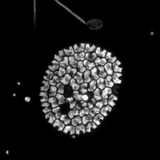
The natural fluorescence of the algae can be observed as the confocal microscope scans through the different planes (height) of the water sample. The image on bottom left is the b/w image of Pediastrum sp. integrated over the Z axis of the colony.
The picture to the bottom right is a different species of Pediastrum.
The picture to the bottom right is a different species of Pediastrum.
View in Media Gallery
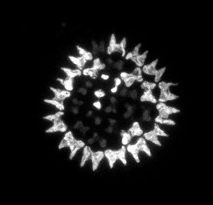
Colony of alga as seen with a confocal microscope
View in Media Gallery
Another species of algae as seen under the confocal microscope
View in Media Gallery
An algal colony organises in the shape of a hollow sphere as it floats in water. Can you identify the species?
View in Media Gallery
Rib-like structures suspected to be diatoms can be also seen under the confocal microscope.
It was encouraging and humbling to learn that the Foldscope can examine a drop of water quite comparably to state-of-art microscopes used for advanced scientific research!
See more about the fauna of Dal Lake in the next post – Part II 🙂
Cheers,
Ashalatha
– with Chandrika and Purnati
It was encouraging and humbling to learn that the Foldscope can examine a drop of water quite comparably to state-of-art microscopes used for advanced scientific research!
See more about the fauna of Dal Lake in the next post – Part II 🙂
Cheers,
Ashalatha
– with Chandrika and Purnati
Sign in to commentNobody has commented yet... Share your thoughts with the author and start the discussion!

 0 Applause
 0 Comments