Foldscope Workshop in Trichy, India – Student Reports
 Aug 07, 2016 • 2:59 AM UTC
 Unknown Location
 140x Magnification
 Unknown
Keerthi and Reethi P
Learn about the author...
4posts
6comments
1locations
View in Media Gallery
Note: This post is continuing from
Foldscope Workshop in Trichy, India – DC Micronauts
which describes in detail the workshop that was conducted a couple days ago.
Each and every student that attended the workshop completed a report on what they observed under the Foldscope, either of a sample they made or a pre-prepared slide. Here are some highlights of the reports, from the students who creatively described what they saw.


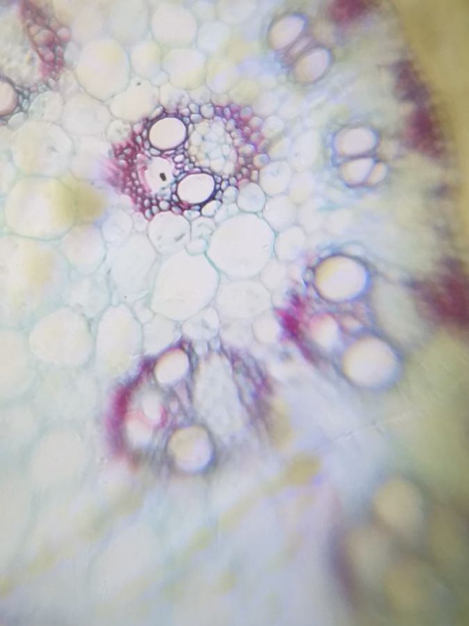
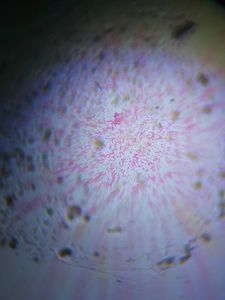
The corn stem, as captured under the Foldscope In this report, P. Suganya and M.Sivasankari did a fantastic report on a corn stem (monocot stem). They creatively compared the vascular bundles to skeletal faces. According to the students,
“They look like a skeleton skull.”
“They look like a skeleton skull.”

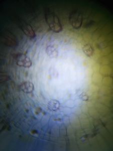
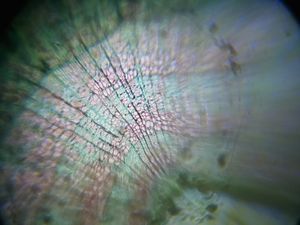
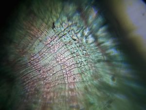
Another view of the corn stem In another report on a cross section of a corn stem by S. Ragul and S. Albert Edison, the corn stem was observed under low magnification and drawn nicely. The students noted,
“It looks like a group of rings!”
“It looks like a group of rings!”

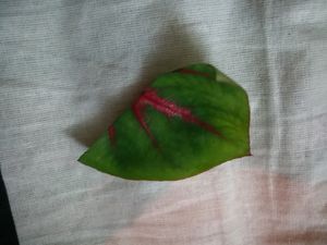
A sample of the caladium leaf
Some images of the pink and green sections of the leaf One student, S. Hariharan viewed a sample of a caladium leaf, a plant with bright pink and green leaves.
“I’m very surprised to look at the cells under the microscope. The leaf looks more beautiful with various colors!”
“I’m very surprised to look at the cells under the microscope. The leaf looks more beautiful with various colors!”

Student S. Gene Albert witnessed a sample of cardiac muscle under the Foldscope and was very impressed by what he saw.
“It looks like strips of black and white in an alternate manner…When I saw this I got an idea of using this for my educational site. Thanking you all for this wonderful opportunity”
“It looks like strips of black and white in an alternate manner…When I saw this I got an idea of using this for my educational site. Thanking you all for this wonderful opportunity”

Student J. Josphin Jenifa commented on the observation of Human Blood Smear under the Foldscope and was thrilled to learn about her sample
“I could see what I could not see through the naked eye. Through this, we learned a lot. Many thanks.”
“I could see what I could not see through the naked eye. Through this, we learned a lot. Many thanks.”

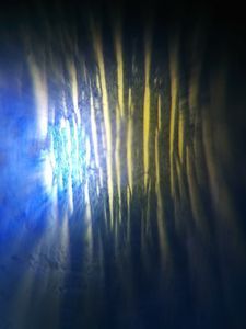
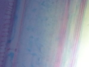
A few images of a Basswood Stem For T. Veera Sethu Raaman and P. Sagaya Nelson, observing the Longitudinal Section of a Basswood Stem was very fascinating. According to Veera and Sagaya,
“I can see many colors like green, red, pink, etc…I was so excited with this Foldscope!”
“I can see many colors like green, red, pink, etc…I was so excited with this Foldscope!”

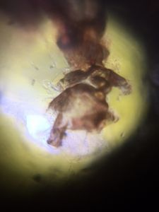
A curcurbita stem Roshan Kumar R. and Logesh R. commented on viewing a cucurbita stem. They went into detail, stating
“It looks like the stem of a tree grounded in the soil…The Foldscope was done by us which excites us to a greater extent!”
“It looks like the stem of a tree grounded in the soil…The Foldscope was done by us which excites us to a greater extent!”

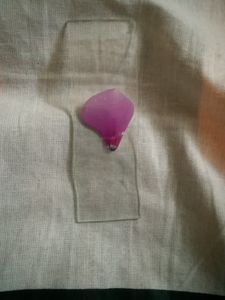
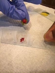
A slide of the nerium flower
View in Media Gallery
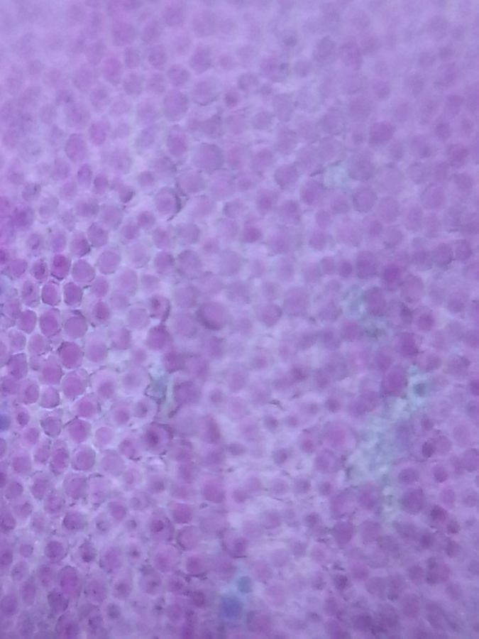
Cells of the purple nerium petal T. Kowshika and A. Zarafath Begum were so dexterous that they were able to observe three specimens in a short period of time! They not only visited the pre-prepared slides section but also made their own sample of the nerium flower petal and a pine leaf (not pictured), and drew and diagrammed what they saw.
“It is tinier than a dot, but in the microscope it is a large, blue circle. We could not see all these tiny particles with our own eyes. “
These, along with many other reports, exemplify how the Foldscope can expand these young minds’ curiosity and spark their creativity imagination. Hopefully, our workshop has ignited the interest of these students so that they continue to observe and question wonderful things within our amazing world!
Sign in to commentNobody has commented yet... Share your thoughts with the author and start the discussion!
 0 Applause
 0 Comments