Life in an ‘Organic pond’ – Part 1
 Oct 28, 2019 • 4:29 AM UTC
 Unknown Location
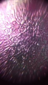
 140x Magnification
 Microorganisms
Jayashree Ramadas
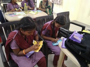
We are a group of students, volunteers and staff working with TIFR Hyderabad's Science Education and Outreach program: http://www.tifrh.res.in/~outreach/
39posts
26comments
2locations
View in Media Gallery
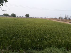
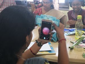
After my experience and learning with the Dal Lake water sample, I have been constantly on the lookout for water ecosystems. Recently, I visited my sister’s home at the ‘Organo Eco-Habitat’ in Hyderabad, where there is an artificial pond.
View in Media Gallery
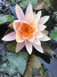
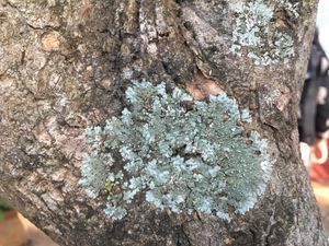
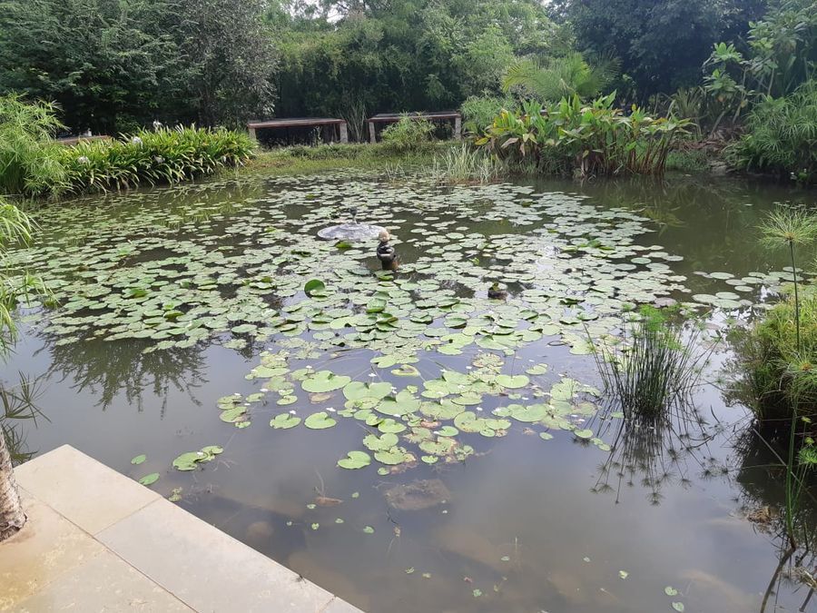
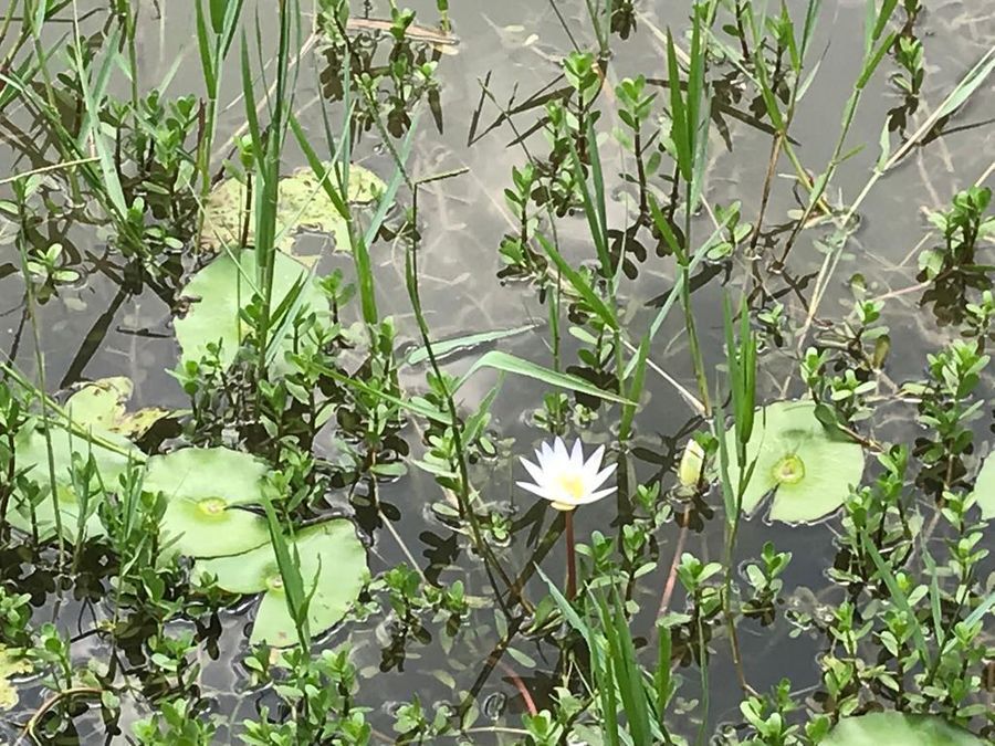
Organo pond with Macrophytes
View in Media Gallery
I collected water from a corner of the pond, in which I found an insect wing to begin with!
I. Life around an insect wing
I. Life around an insect wing
View in Media Gallery
Viewing the insect wing under the Foldscope, I saw many green ciliates with cirri at the posterior end. Are they Euplotes ? And do you see food vacuoles inside the Euplotes ?
View in Media Gallery
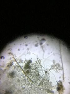
Watch this colourless elongated unicellular animal with a long proboscis at the anterior end and a small tail-like extension in the posterior. We see many vacuoles in the cell body. This animal repeatedly extends its proboscis to gather up microbes. It is a Dileptus sp.
View in Media Gallery
I found this new (for me) species of Rotifer busy scraping the algae attached on the insect wing.
View in Media Gallery
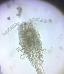
Here is an arthropod with transparent body, a pair of antennae and one red eye. I think it is a Copepod .
II. Life around a decaying leaf
II. Life around a decaying leaf
View in Media Gallery
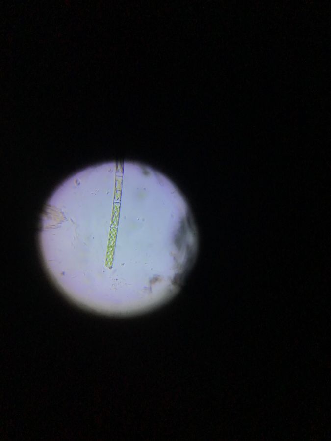
The water looked very clear, but there was a decaying leaf in it. On an impulse I scratched the leaf gently and put the scratched stuff on the slide with a drop of water. A beautiful Spirogyra sp. with spiral chloroplasts came into focus.
View in Media Gallery
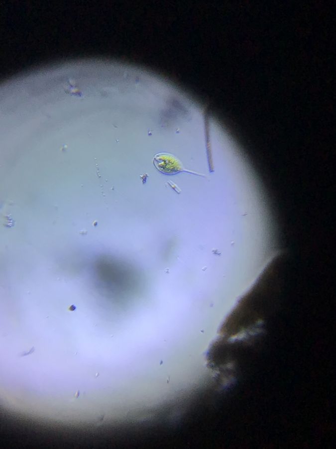
There were different species of Euglenoids in this water sample. Here you see two types of eukaryotes with green pigment and red eye-spots –
one pear-shaped, Euglena sp ., the other leaf-like, Phacus sp ..
one pear-shaped, Euglena sp ., the other leaf-like, Phacus sp ..
View in Media Gallery
The euglenoid locomotion of this long tailed green eukaryote is mesmerizing. This beauty is a genius in physics and biology!! It is Phacus helicoides .
View in Media Gallery
Phacus are not commonly found in stagnant water due to lack of sufficient organic deposition. Since different species of Phacus are present in this pond, it means there is rich organic matter around.
Here is another euglenoid, to the left of a diatom. See how it slowly stretches out its body! I think it is Euglena ehrenbergii.
Swimming around the periphery of the field of view is another pear-shaped Euglena sp..
Here is another euglenoid, to the left of a diatom. See how it slowly stretches out its body! I think it is Euglena ehrenbergii.
Swimming around the periphery of the field of view is another pear-shaped Euglena sp..
View in Media Gallery
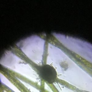
Here is a Heliozoan , commonly called a ‘sun – animalcule’, which is a protozoan with many axopodia arranged radially. We can clearly see the ectoplasm, endoplasm and a green food vacuole to its right.
III. All in one drop!
III. All in one drop!
View in Media Gallery
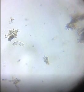
There was a sudden jerk in the slide. As I focused I saw two worms moving swiftly.
View in Media Gallery
The anterior end looks like a head with two eyes and the posterior end has hair.
A mosquito larva would have hair/bristles all over the body, so I am certain this ‘worm’ is something else.
A mosquito larva would have hair/bristles all over the body, so I am certain this ‘worm’ is something else.
View in Media Gallery
I searched a lot to identify the species and came to the conclusion that it could be a Chironomid larva. Can anyone verify this?
View in Media Gallery
I found another worm which is segmented and has bristles on either side of its transparent body. Its intestine can be seen too. I think this is an annelid, Chaetogaster sp ..
View in Media Gallery
By this time, I had been working on the sample for three to four days. I found many Phacus sp . dead 🙁 Perhaps, it was because of lack of organic matter.
IV. Life in green mass
IV. Life in green mass
View in Media Gallery
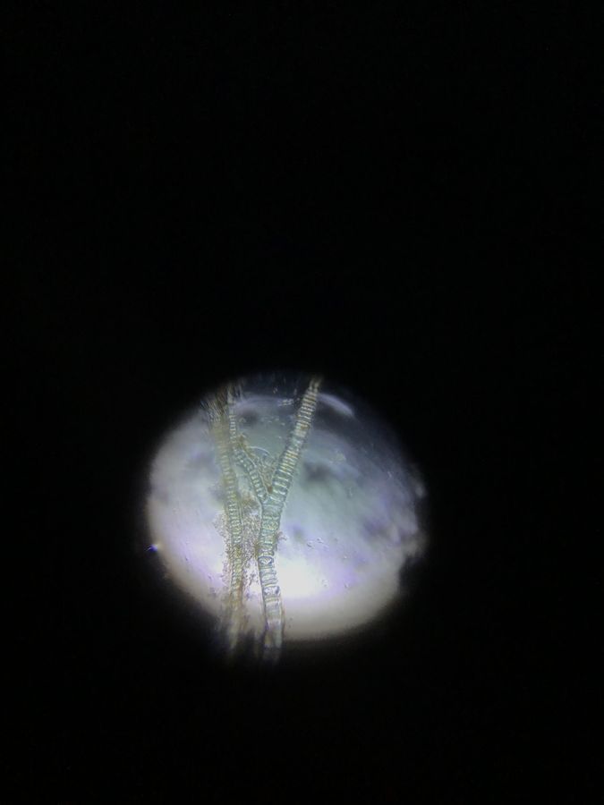
I had collected some green mass separately from the pond. This slide was very special as it offered such a colourful experience. Watch the different types of Diatoms disperse the focussed light.
I love this video, such beauty in nature!
I love this video, such beauty in nature!
View in Media Gallery
This dark green mass is surely branched Cyanobacteria .
View in Media Gallery
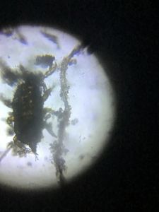
I seem to have disturbed a water flea while it was busy foraging.
View in Media Gallery
I found an Ostracod . I had to wait a long time for it to open its bivalve shell and so that its appendages could be seen to confirm. It needed all the patience I could muster and it was totally worth it!
Check out some amazing facts about Ostracods from the Lake Biwa Museum in Japan.
I had such an enriching experience with this water sample, but it saddens me that in the process of these discoveries many of my microbe friends were killed, especially Phacus helicoides and the beautiful diatoms ! I understand this is one outcome of our human curiosity and learning and yet, going ahead, I do hope to find new microbe friends in the waters around India. Our journey with the Foldscope continues…
Cheers,
Ashalatha
with Chandrika and Jayashree
Sign in to commentNobody has commented yet... Share your thoughts with the author and start the discussion!

 0 Applause
 0 Comments