Mysteries of Dal Lake, Kashmir – Part 2
 Oct 16, 2019 • 6:52 AM UTC
 Unknown Location
 140x Magnification
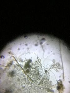
 Microorganisms
Jayashree Ramadas
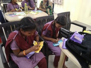
We are a group of students, volunteers and staff working with TIFR Hyderabad's Science Education and Outreach program: http://www.tifrh.res.in/~outreach/
39posts
26comments
2locations

View in Media Gallery
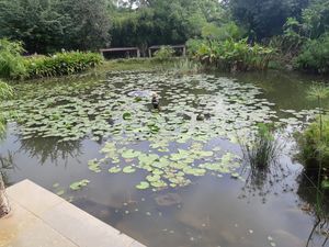
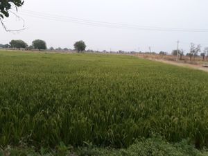
After seeing the diverse flora in Part I , here I share the findings on the microbes in the water from Dal Lake.
View in Media Gallery
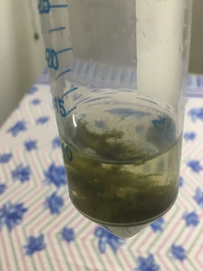
We thought we could find a dense composition of organisms if the water could be centrifuged. Sayantan, a graduate student in biology at TIFR Hyd helped me with this. We put 50 ml of lake water in a centrifuge chamber at 25°C, 500 rpm for 3 minutes.
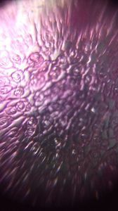
It is interesting to see the dynamics of these microbes around algae. That is the key to finding organisms in a water sample – look for them around algal matter!
It is interesting to see the dynamics of these microbes around algae. That is the key to finding organisms in a water sample – look for them around algal matter!
View in Media Gallery
Active microbes moving around an algal filament with diatoms floating around.
View in Media Gallery
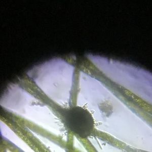
Here is a ciliate foraging – Euplotes sp. The rib-like and star-like diatoms can also be seen. A rotifer (in a different plane) makes a special appearance!
View in Media Gallery
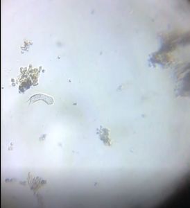
Here is a lone and swift moving Rotifer as seen under the Foldscope.
View in Media Gallery
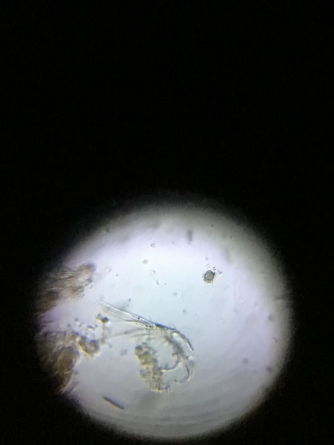
Here is a Rotifer practicing gymnastics around filaments of green algae. (I think the algae is Cladophora sp .. Is it correct?)
To the right of the field of view, an organism like a
Crustacean can be seen.
To the right of the field of view, an organism like a
Crustacean can be seen.
View in Media Gallery
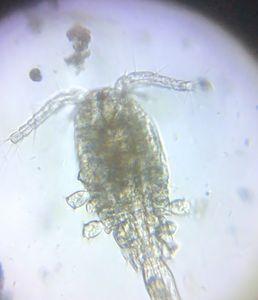
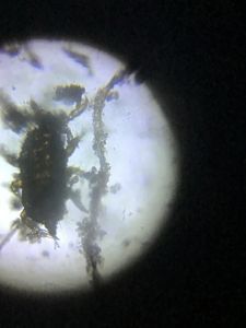
Here is a closer look at the
Arthropod. Can anyone identify the species?
Arthropod. Can anyone identify the species?
View in Media Gallery
Here we can see different species of rotifers. While one is active the others seem to be quietly feeding under the shade of algae 🙂
After a few weeks of getting the water sample, I noticed a sudden increase in activity of its microorganisms.
After a few weeks of getting the water sample, I noticed a sudden increase in activity of its microorganisms.
View in Media Gallery
These look like hundreds of ciliates swiftly moving in the water. I wonder what triggered this population explosion?
View in Media Gallery
I observed the same slide 12 hours later and saw this – I was not able to identify these fast moving microbes!
View in Media Gallery
Are they ciliates or young rotifers? They seem to be dominating the water sample at that moment.
View in Media Gallery
See the redish rotifers in the background. Would they be dormant or dead? Rotifers feed on dead bacteria, protozoans and algae, so I thought increase in ciliates in the sample would signal happy times for Rotifers ! However, I am not sure what is happening here!?
View in Media Gallery
And finally, here we can see the locomotion on an Amoeba . This was captured under the confocal microscope at 100x magnification – imaged by Purnati, TIFR Hyd.
I was very surprised that ever after two months of sample collection (about 750 ml of water was kept in a water bottle with the cap open), there was a rich ecosystem. No matter how many times I went back to study the sample, I always found something novel and exciting! I spent the summer and monsoon months examining this sample and now am hooked on to observe the beautiful micro-cosmos in a drop!
Cheers,
Ashalatha and Chandrika
– with thanks to Sayantan and Purnati
Sign in to commentNobody has commented yet... Share your thoughts with the author and start the discussion!

 0 Applause
 0 Comments