Ganges River microbial diversity
 Aug 21, 2016 • 7:25 PM UTC
 Unknown Location
 140x Magnification
 Microorganisms
Saad Bhamla
Learn about the author...
32posts
11comments
2locations

View in Media Gallery
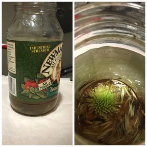
I received a strange message from a colleague at Stanford (Julie F.). She reached out to me via Facebook and said that a friend of hers (Kathy H.) had returned from a Fulbright scholarship in India and had brought a bottle of Ganges River water with her, obtained from Varanasi. Kat is a teacher at the Packard Children’s Hospital School at Stanford and wanted to share her experiences with kids who are admitted into the hospital for long treatment periods and often have to miss school. So, long story short, Julie came walked over to my office and handed me a bottle of brown muddy water, which I of course offered to foldscope – I couldn’t believe my fortune – a bottle of precious biological specimen that had flown all the way across the Pacific – it felt like Christmas had come early this year:)
I was worried though. The bottle had been sealed for a few days and I was concerned that lack of oxygen could have killed off most of the microorganisms (Manu assured me that the mixing during handling and travel would have kept it aerated, and he was right,as you will find see). I transferred it into a flask for aeration and left it overnight before sampling.
I was worried though. The bottle had been sealed for a few days and I was concerned that lack of oxygen could have killed off most of the microorganisms (Manu assured me that the mixing during handling and travel would have kept it aerated, and he was right,as you will find see). I transferred it into a flask for aeration and left it overnight before sampling.

View in Media Gallery
Here’s a close-up of the bottom.

View in Media Gallery
So I took a small drop from the bottom of the flask, prepared a wet mount and loaded into the foldscope.
Let’s dive in. In the video below you’ll see (and hear) my astonishment at finding plenty of life – ciliates, bacteria, and a swimming Euglena.
Let’s dive in. In the video below you’ll see (and hear) my astonishment at finding plenty of life – ciliates, bacteria, and a swimming Euglena.
So that answered the question of whether there were living things still in the sample. In the next video, I stayed focused at one place to follow the trajectories of two strange swimmers – one vibrates (shivers) erratically and the does a drunken walk..
I decided to revisit the Euglena, as it seemed to be the dinosaur in my drop. And I had never seen one swim before. As you watch below, notice how it uses its flagella to pull itself – often flagella are used as propellers to push, but to my amazement, here the Euglena pulls itself. How does it do that? How do you take a flexible object and use it to pull yourself? I’m imagining a rope that the Euglena spins (observe the fine vibrations) that create a net force to propel itself. It just doesn’t make any sense to me.. How is this possible?
Curious to what else was in the water, I created a second slide with a fresh drop of water from the flask. I made sure to mix it this time to pick up as many organisms as I could. In this next video, watch as I observe a huge beast look lurking in the shadows (but doesn’t reveal itself), an ultra-fast ciliate and a beautiful square shaped diatom.
And then as I was scanning, something caught my eye. An extraordinary contraction – so sudden that I missed it at first. But as I peeked closely, I saw a flow remnant of ciliates that create small flows for sucking bacteria and other tiny cells to their mouths. As I watched, the ciliate spontaneously contracted again! What a beautiful sight. How does it do that? Why does it do that? Does it have a spring inside? Imagine if moved that rapidly- wouldn’t your internal organs get damaged- how does this ciliate ensure it doesn’t pass shocks through its own body?
The next video is a brief one in which a green spiky object caught my attention – what is it? Could it be a microscopic leaf? Or a diatom? Or an algae?
For my last video, to share with the kids who Kat might show these videos to, I made it easy to visualize the hundreds of millions of bacteria in the water by using the dark field mode of the foldscope. Watch the whole video to enjoy the blinking lights of tiny bacteria waving their lanterns at you, saying hello, all the way from Varanasi, India.
Thanks again Kat and Julie for sharing this previous water samples.
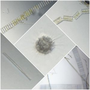
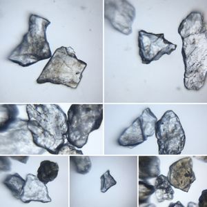
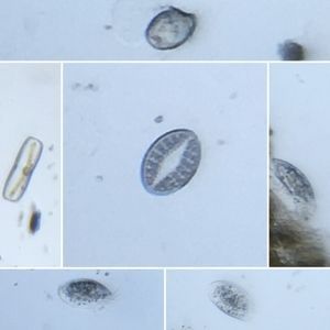
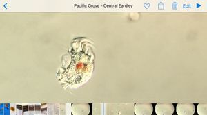
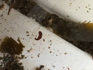
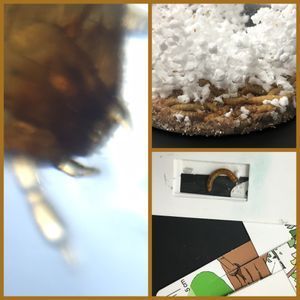
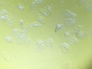
These are a few images from the samples.
These are a few images from the samples.


Sign in to commentNobody has commented yet... Share your thoughts with the author and start the discussion!

 0 Applause
 0 Comments