Foldscope excitement at ZPHS Vattinagulapally
 Nov 18, 2019 • 8:41 AM UTC
 Unknown Location
 140x Magnification
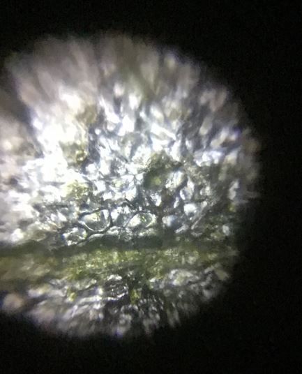
 Microorganisms
Jayashree Ramadas
We are a group of students, volunteers and staff working with TIFR Hyderabad's Science Education and Outreach program: http://www.tifrh.res.in/~outreach/
39posts
26comments
2locations
View in Media Gallery
Zilla Parishad High School (ZPHS) Vattinagulapally is a semi-rural local government school on the outer fringes of the city of Hyderabad, Telangana. I have been visiting this school as a volunteer in the weekend outreach program of TIFR Hyderabad, since July this year.
Classes at Vattinagulapally are fairly small, partly because of drop-outs after elementary school and partly due to the flight of students from government to private schools nearby. In Class 8 there are 23 students on the rolls, 10 girls and 13 boys. The school follows the State approved curriculum in Telugu medium. The bioscience textbook for Class 8 opens with the ‘scientific method’ (a wonderful backdrop for foldscopes!), moving on to the introduction of cells, unicellular and multicellular organisms, shapes and sizes of cells, and so on.
Between end July and early September 2019 I taught four classes to students of Class 8, to introduce them to the microscopic living world. In the first visit I showed them some Foldscope videos taken earlier on my iPad – Spirogyra and Vorticella , which happen to be pictured in their textbooks, and then some videos of nematodes and rotifers .
View in Media Gallery
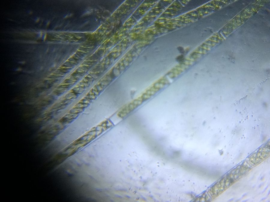
Spirogyra in pond water.
The students got very excited and immediately brought out their textbooks, searching them for figures and information about micro life-forms.
The students got very excited and immediately brought out their textbooks, searching them for figures and information about micro life-forms.
View in Media Gallery
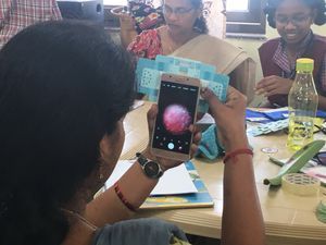
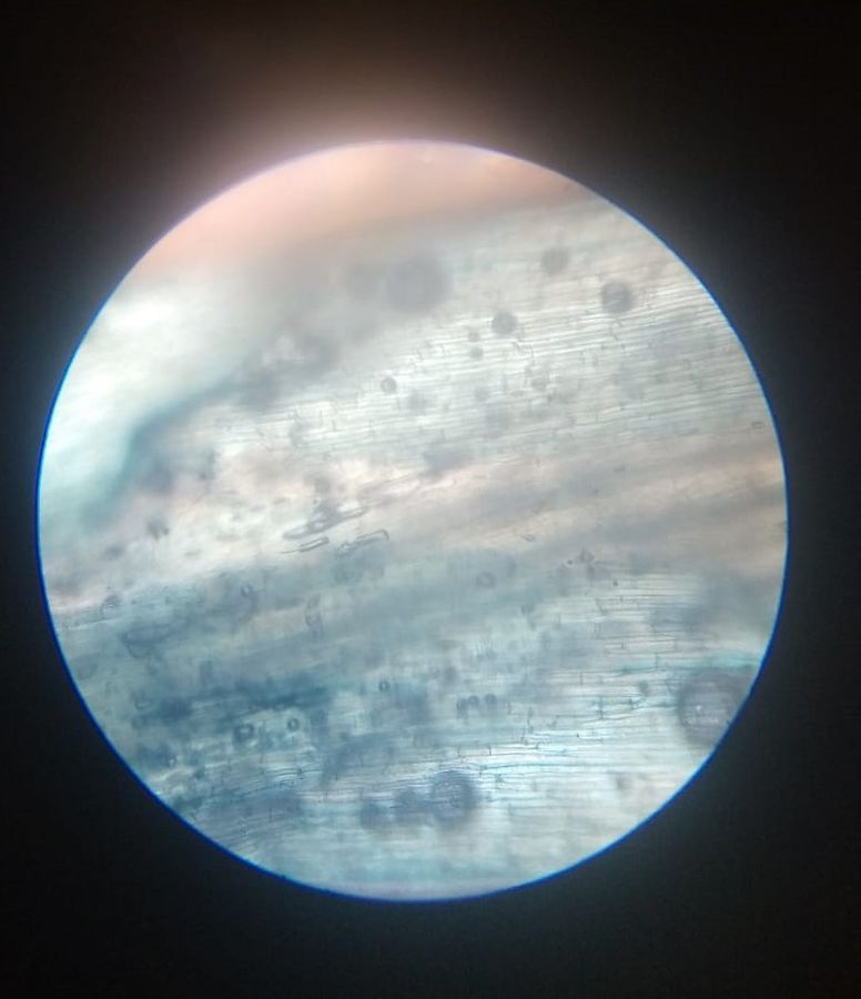
Then we brought in the compound microscope from their school lab, and students got their very first experience of making and viewing slides of a leaf epidermis (perhaps it was Ixora) and observing them under a compound microscope. Later I showed them the same slides under my Foldscope. They were enthusiastic and captured these images on my iPad.
View in Media Gallery
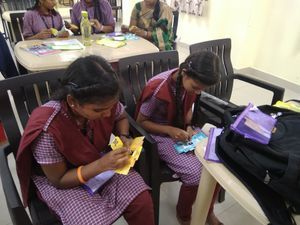
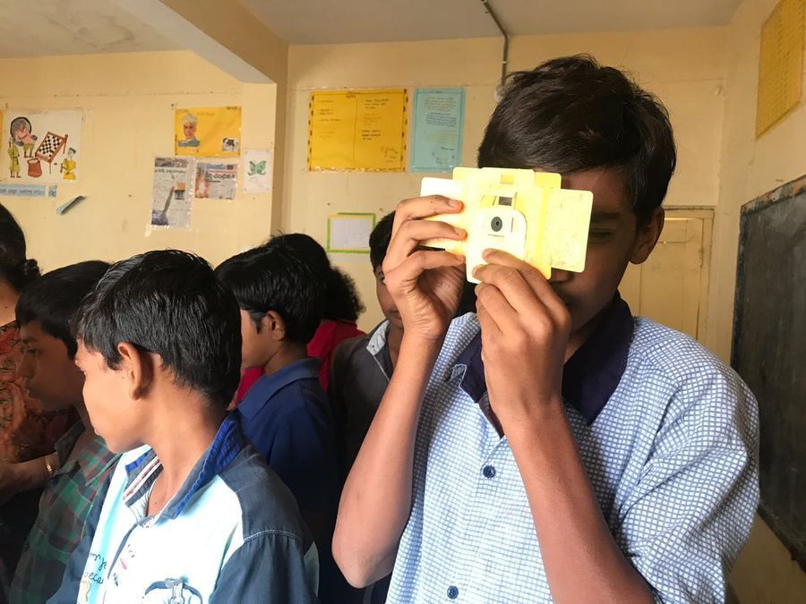
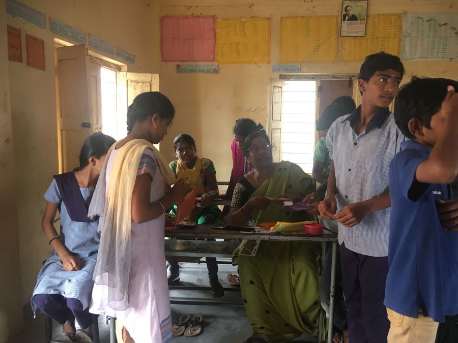
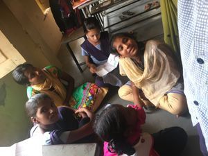
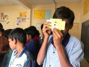
In the next visit I went with three new Foldscope kits. I gave one kit to a group of five boys, one to five girls and the third to their biology teacher Ms. Shanthakumari. They followed the instructions and folded them in half an hour.
The students and their teacher were very excited while focusing the slides made by them in their own folded Foldscopes!
The students and their teacher were very excited while focusing the slides made by them in their own folded Foldscopes!
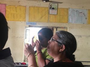
Driven by that excitement, they made different slides of monocot leaf epidermis, meristematic tissue, pollen grains, etc.. They also took a video of their observation of an ant’s leg.

View in Media Gallery
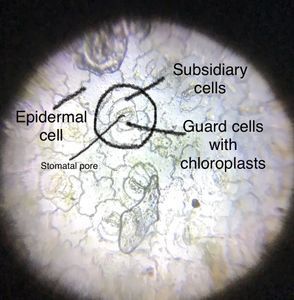
Stomata in lower epidermis of Philodendron

View in Media Gallery
Philodendron plant

View in Media Gallery
Lower epidermis of a monocot leaf
View in Media Gallery
Meristematic tissue of roots of an onion plant.

View in Media Gallery
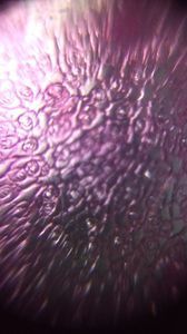
Pollen grains
View in Media Gallery
Ant leg under the Foldscope
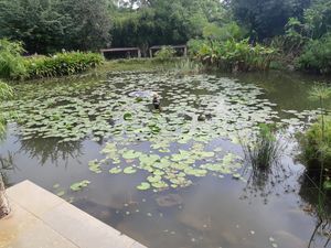
In the third visit I took with me some water from our Dal Lake water sample (see the posts 1 & 2 ) and taught the students how to prepare a wet mount. The teacher was now fully enthused and the following Saturday (a school holiday) she collected some rain water from around her house and landed up at my home, where we spent a pleasant afternoon observing a rotifer colony.
In the third visit I took with me some water from our Dal Lake water sample (see the posts 1 & 2 ) and taught the students how to prepare a wet mount. The teacher was now fully enthused and the following Saturday (a school holiday) she collected some rain water from around her house and landed up at my home, where we spent a pleasant afternoon observing a rotifer colony.
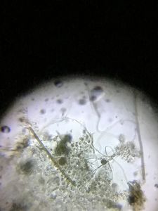
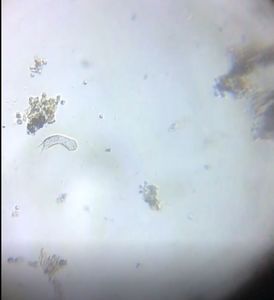
View in Media Gallery
Here is a colony of rotifers in rain water – wet mount prepared by Ms. Shanthakumari.
View in Media Gallery
Back in school, from this same sample, a video of a rotifer’s locomotion was prepared and observed by ZPHS students.
View in Media Gallery
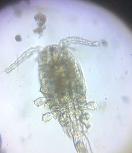
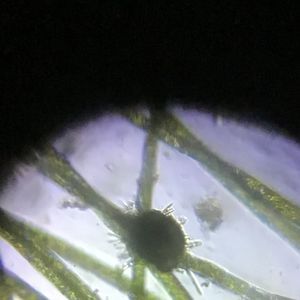
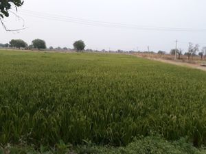
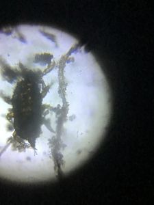
This event further inspired the students, who collected stagnant rain water near the school, prepared a perfect slide and showed it off in my next visit to the school. Wow! It is Daphnia!
In the days that followed Ms. Shanthakumari and her students shared their experiences with the students and teachers of other classes too. Such active learning, in so many classes of Vattinagulapally – all with just three foldscopes!
Foldscopes are excellent learning and teaching tools for high school students, who are delighted to see the microscopic structures of plants and animals that they have only known as textbook figures, or occasionally as permanent slides. But now, after two years of activity the outreach group is running out of foldscopes! Is someone listening out there?
Cheers,
Ashalatha
with Chandrika and Jayashree
Sign in to commentNobody has commented yet... Share your thoughts with the author and start the discussion!

 0 Applause
 0 Comments