The Power of Concentration
 Sep 07, 2016 • 1:45 PM UTC
 Unknown Location
 140x Magnification
 Microorganisms
Max Coyle
Learn about the author...
16posts
17comments
3locations
View in Media Gallery
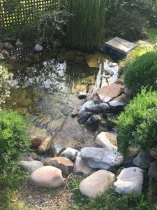
My favorite Foldscope experiment is simple: collect pond or lake water and find out what is inside. It is perhaps THE classic microscopy experiment since the days of Leeuwenhoek. Discovering the strange life inside of a single drop of water is always satisfying.
I recently visited Lake Anza in Berkeley’s Tilden Park to collect water and characterize the micro-biodiversity of the place. A drop of lake water under the Foldscope allows you to see much – algae, ciliates, strange types of debris..but you also tend to see the same, most common forms over and over again.
To find a wider diversity of life, I decided to try concentrating my samples. I used a nylon mesh with a 5 micron pore size, folded in into a funnel, and passed maybe 100 mL of lake water through. The 5 micron filter size should allow bacteria to pass through, but should trap many larger single-celled eukaryotes and small metazoa. When the sample had almost all the way passed through the filter, so there was only a few drops remaining in the cone, I collected one of these drops for the Foldscope.

View in Media Gallery
Nylon mesh (5 micron pore size) folded into funnel Within my concentrated sample, I saw an abundance of interesting life forms…
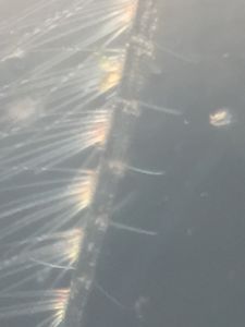
1)First was this cell with rapidly beating cilia all around it:
1)First was this cell with rapidly beating cilia all around it:
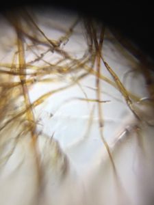
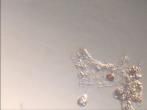
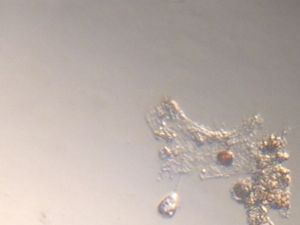
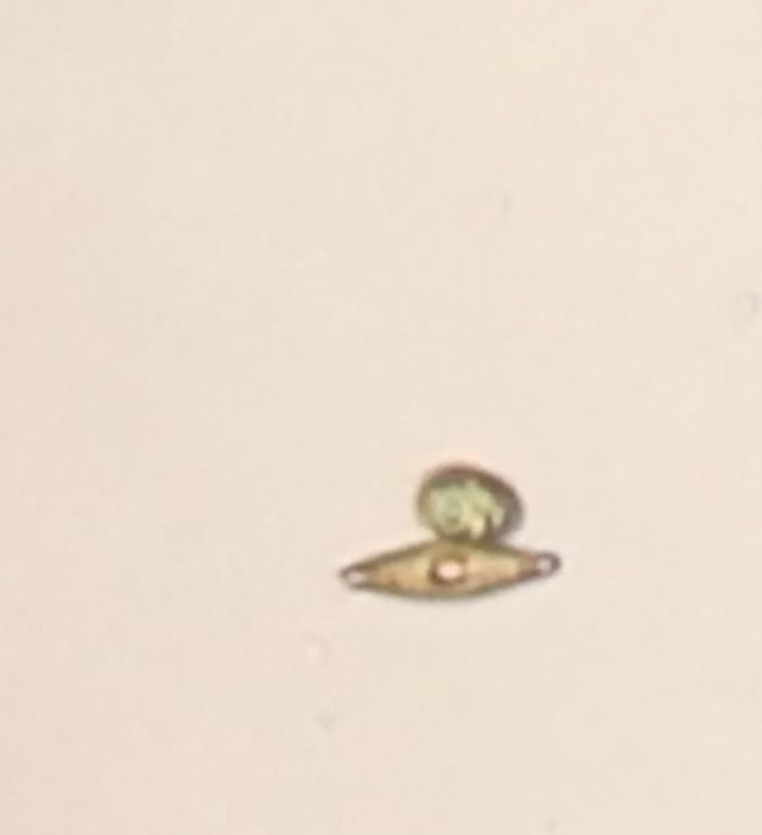
2) Next, I found a pair of interesting creatures interacting with one another: an orange-ish diatom and a green algae. I was fascinated observing them and condensed all my footage down into the following clips, which show the pair moving together, as well as each species moving individually. They are not irreversibly anchored, as the point of attachment varies, and as you will see towards the end, they eventually separate temporarily, re-connect, and then separate entirely. I do not know if this interaction is rare or not, and am unsure of its purpose.
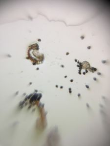
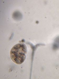
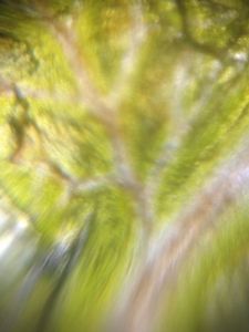
3) I saw this cluster of algae, with two different shades of green, a darker one and a paler one. Is it two different species or variety within the same species?
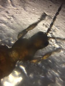
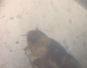
4) Finally, I was fascinated by this rapidly flailing creature. At first I thought it was the same ciliate as 1), but then realized that the motions were not 360 degrees around, but were localized to one side. Also, the appendages appear thicker than cilia, making me wonder if this was some kind of larval form.
As always, your thoughts, observations, and identifications are welcomed!
Until next time,
Max
Until next time,
Max
Sign in to commentNobody has commented yet... Share your thoughts with the author and start the discussion!
 0 Applause
 0 Comments