The all-engulfing Sun: Experiments with the heliozoan Actinosphaerium
 Dec 31, 2019 • 12:13 PM UTC
 Unknown Location
 140x Magnification
 Microorganisms
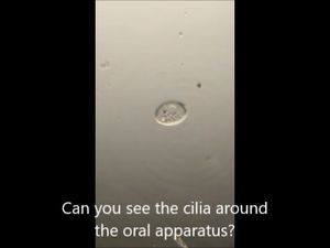
Laks Iyer
Human observer of life. https://sukshmadarshin.wordpress.com
97posts
1255comments
5locations

View in Media Gallery
Actinosphaerium, also called a helizoan or sun-animalcule, belongs to the SAR- (Stramenopile-Alveolate-Rhizaria) branch of the eukaryotic tree. Recent phylogenetic analyses suggest that they are closer to haptophytes in the tree of life [ To access a formal study on the matter, click here ]. These “Sun-shaped” eukaryotes have stiff needle-like pseudopodia (also called axopods/axopodia) that surround their spherical body. I have for long been wanting to study them and got a delightful opportunity recently. This sample is a commercial sample from Carolina Biologicals, but I have often found them in lake/pond water during the summers. They are pretty easy to grow in boiled-wheat medium with a prey such as Paramecium tetraurelia. In general, they are non-fussy and also grow well when provided rotifers.
Basic anatomy . Three basic components can be delineated for the helizoans. Watch the next video.
Basic anatomy . Three basic components can be delineated for the helizoans. Watch the next video.
2. Avoidance on contact with axopods One striking aspect is the recoiling of ciliates and other critters on contact with the Actinosphaerium axopods. In the following video, notice how Paramecium and Bursaria avoid contacting the pointy tips of the heliozoan axopodia. What might the cause of this aversion? Are the tips of the pseudopods tipped with a toxin that is injected as speculated, or is there some kind of electric potential discharge, or is it just pin-tip pressure? Recent studies indicate that Ca-ion concentration fluxes affect the assembly and disassembly of axopods. Could these be injecting some ions depolarizing other ciliates? Detailed physiological studies are few for the heliozoans, or perhaps very old, and these are ripe for a revisit with modern imaging tools and reagents.
2. Predator-prey interaction . While many critters have this aversion response on contacting the axopods, some are not so fortunate and meet their end in the spears and are then engulfed by the heliozoan center.
3. The process of phagocytosis . The above motivated me to document the process of eating more thoroughly. Timelapse photography is a productive method of doing this and studying heliozoans in general. For this, I used the Lapseit app on my phone. Some of these timelapse movies were recorded overnight: I kept the foldscope right next to where I sleep to make adjustments if the organism went past the field of view. It is fascinating to see how prey pierced by the Actinosphaerium is taken up into the endoplasm. It is almost as though the spines melt away to bring the prey closer to the center. The key molecular component involved in this process are microtubules that assemble and disassemble based on the requirements. The process is fascinating to watch.
Here is a video with multiple ingestion events. Also, study the axopods “melting away”.
Here is a video with multiple ingestion events. Also, study the axopods “melting away”.
3. Binary fission. Actinosphaerium has at least two different recorded ways of propagation. They often undergo what is called plasmotomy where the eukaryote just breaks off into several different pieces. Binary fission is a special case of the same. The second type of propagation is encystment followed by release of the cyst under favorable conditions to give many progeny forms. Below is a binary fission event. Happy 2020 everyone. Keep foldscoping.
Sign in to commentNobody has commented yet... Share your thoughts with the author and start the discussion!

 0 Applause
 0 Comments
_300x300.jpeg)