Different concentration of K562 and HELA cells under Microscope and Foldscope
 Jan 20, 2020 • 8:38 AM UTC
 Unknown Location
 140x Magnification
 Unknown
annu gowda
Learn about the author...
1posts
0comments
1locations
View in Media Gallery
60ul of HELA cells under Microscope
View in Media Gallery
60ul of HELA under Foldscope
View in Media Gallery
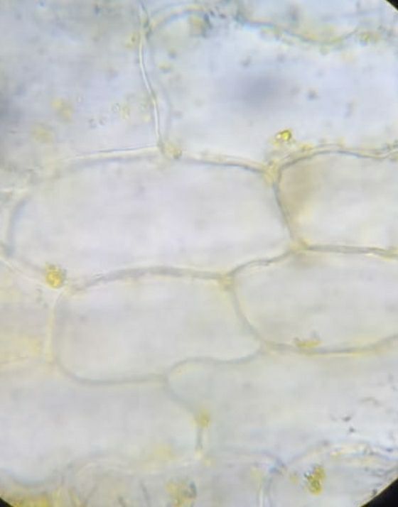
Microscopic view of Marigold flower
View in Media Gallery
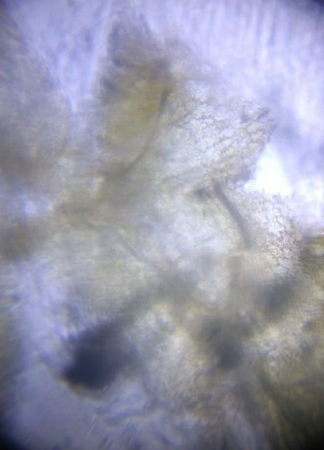
Foldscope view of Marigold flower
View in Media Gallery
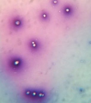
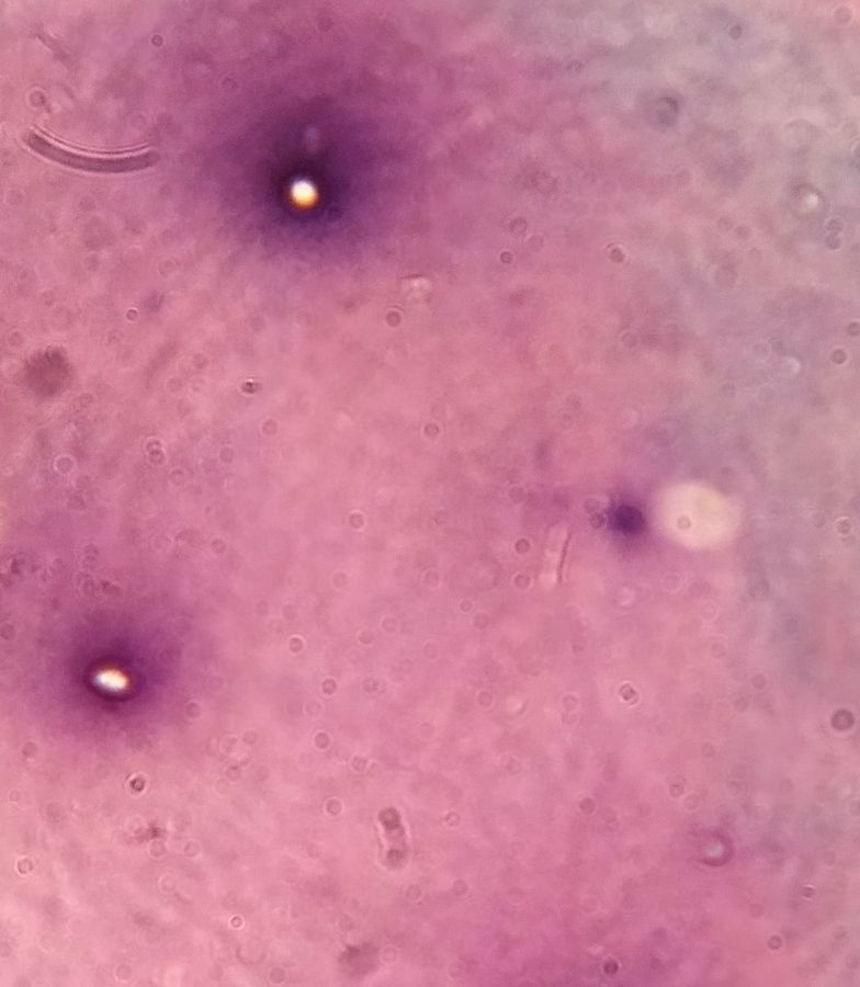
Leishmans stain of HELA cells under Microscope
View in Media Gallery
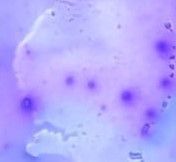
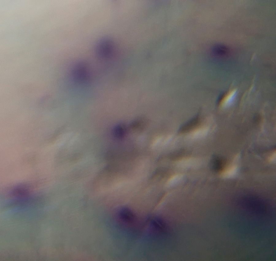
Leishmans stain of Hela cells under Foldscope
View in Media Gallery
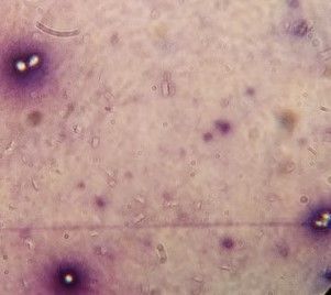
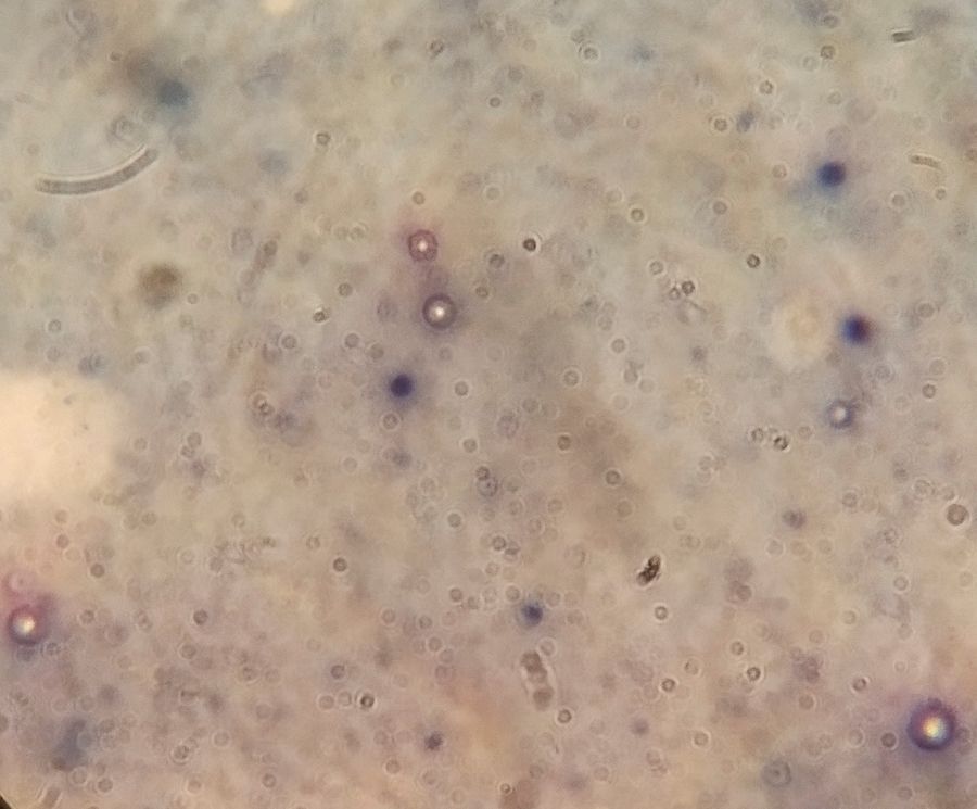
Geimsas stain of Hela cells under Microscope
View in Media Gallery
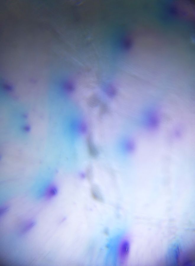
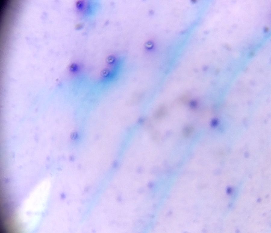
Geimsas stain of Hela cells under Foldscope
View in Media Gallery
Methylene blue stain of Hela cells under Microscope
View in Media Gallery
Methylene blue stain of Hela cells under Foldscope
Sign in to commentNobody has commented yet... Share your thoughts with the author and start the discussion!
More Posts from annu gowda
No more posts from this author.