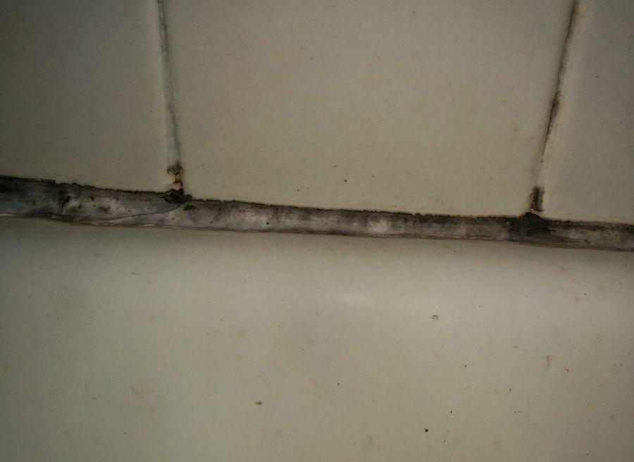
Bathroom mildew
 Feb 16, 2015 • 9:22 PM UTC
 Unknown Location
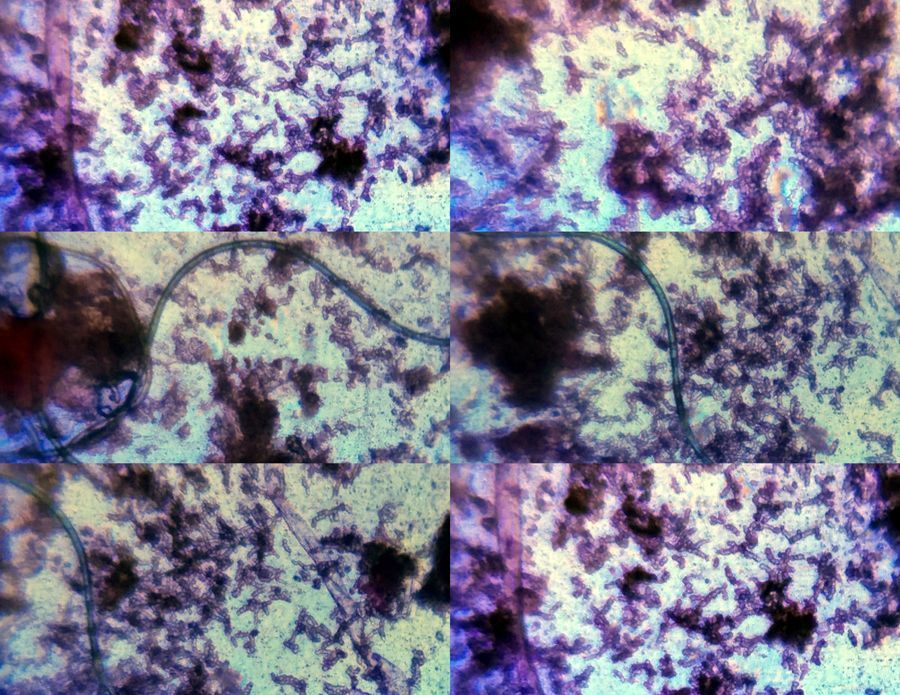
 140x Magnification
 Microorganisms
Laks Iyer
Human observer of life. https://sukshmadarshin.wordpress.com
97posts
1255comments
5locations

View in Media Gallery
I saw some mildew growing in my bathroom caulking. I am calling this a mildew as it isnt woolly or downy. These sticky fellows use the bathroom caulking as substrate and break it down over time. Bleaching them out is the only way to clean the caulking. Since I had the unenviable task of getting rid of them, I thought I’d first foldscope them and study their features.
View in Media Gallery
For this, I took some clear tape and pressed the sticky side on the black midew, to which the fungus stuck easily. I then stuck the tape to a drop of stain on a slide and put it directly under the foldscope. No fixing or washing, easy and elegant. This method (with more specific stains) is even used for fungi that grow on the skin or anywhere for that matter ( Google cellotape flag ). I used 0.5% Carbol Rose Bengal as the stain (after many trials and errors, I found that it stains fungi nicely and it isnt very expensive and an amateur like me can buy it). I like this tape method as it is easy, mess-free, and can easily be viewed using the low and high powers of the foldscope.
The mildew was mainly yeast-like with very few mycelial structures, although the yeast-like structures appear to be in chains. In addition to the pink color of the stain there is a dark pigment of the fungus itself. All these give a nice contrast.
The mildew was mainly yeast-like with very few mycelial structures, although the yeast-like structures appear to be in chains. In addition to the pink color of the stain there is a dark pigment of the fungus itself. All these give a nice contrast.
View in Media Gallery
At higher power the yeast-like branched structure is more prominent. Some hyphal structures were seen, which seemed septate.
View in Media Gallery
My best guess for a name is Aureobasidium, although I am hoping some mycologists take one look at this and tell me what it is, as it is likely to be a common mildew and my mycology is rudimentary. If you have such in your bathrooms, could you foldscope them so that we can see what the diversity of the bathroom mildew is like?
Now for that bleach …
Now for that bleach …
Sign in to commentNobody has commented yet... Share your thoughts with the author and start the discussion!

 0 Applause
 0 Comments
_300x300.jpeg)


