Maggots and Fungi: A Tale of Quarantine time Exploration
 Mar 25, 2020 • 1:36 PM UTC
 Unknown Location
 140x Magnification
 Unknown
Manan Suri
My name is Manan. I am a student from New Delhi, India. I like to explore the world around me, and foldscope acts like a gateway for me to delve into the microcosmos. Always open to learning new things, meeting new people and and having new memories.
16posts
33comments
1locations

View in Media Gallery
Sitting at home has given us a fresh opportunity to look at our homes with a different lens. While rummaging through my garden, I found that my mother had tried to plant a tomato plant in a pot.

View in Media Gallery
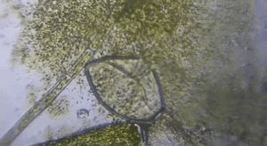
the rotting tomato pulp; black algae can be seen surrounding it The tomato pulp present in the pot was undergoing decay and dessication by fungi, and insect larvae (maggots).
So naturally out of curiosity I carefully collected the samples and made slides, observing them through the foldscope.
The first sample I took was of the fungi. I could see the hyphae- which are thread like filaments which are constitutive of the fungi itself. These are branched and intertwined to allow maximum area for growth and saprophytic action. These hyphae form the mycelium which is the vegetative part of the fungi.

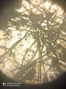
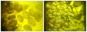
Throughout the slide I could see abundant and very small particles, which are the spores
Furthermore, at the end of the hyphae,I could observe the source of these spores: the fruiting body (sporocarp) from where the spores are released.
These spores fly distances and land on surfaces. Whe suitable conditions for growth of fungi are found, I.E. warm and moist with availability of organic matter, these spores germinate to form a new network of fungi.


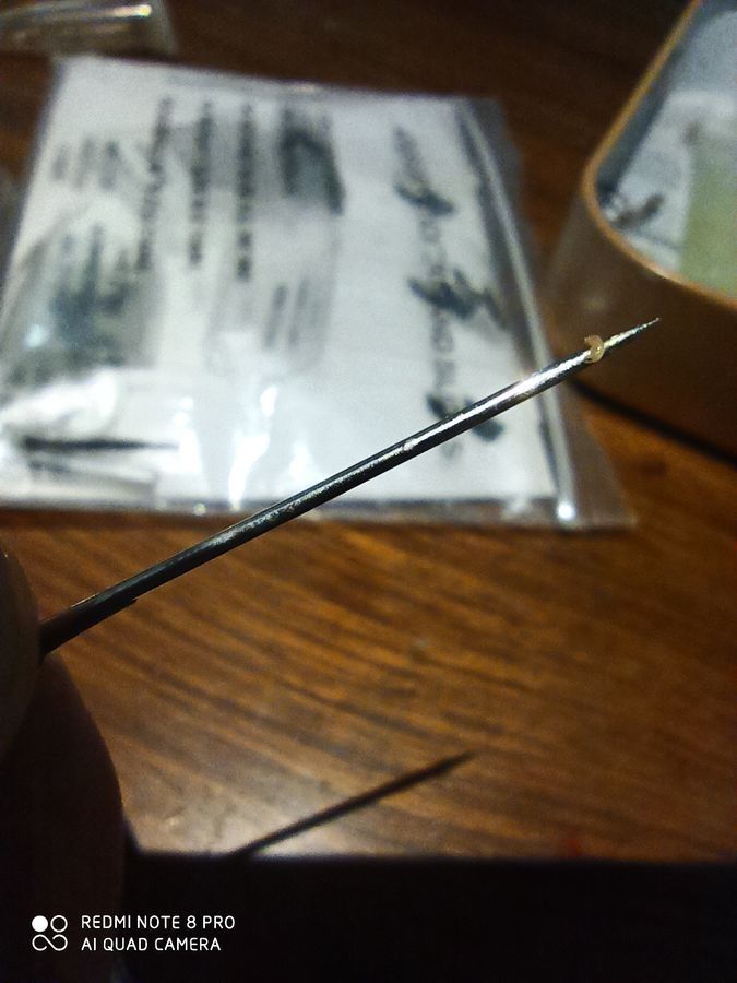
Along with the fungi which are saprophytic, another type of organism that I found was very small maggots/ insect larvae.
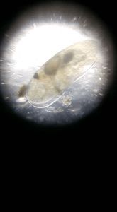
You can see one of them which I picked up on a needle:
You can see one of them which I picked up on a needle:
View in Media Gallery
I carefully used the silicon well slide to form a slide of this tiny creature.
View in Media Gallery
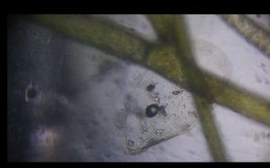
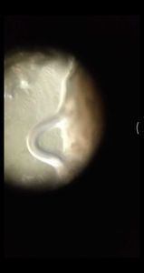
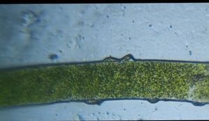
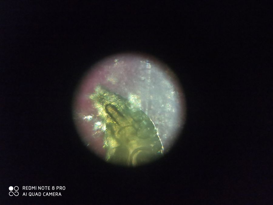
The posterior end of the larva. We can see the tomato pulp it has consumed.
View in Media Gallery
The anterior portion

View in Media Gallery
Anterior portion


We can see the structure of the ‘mouth’ In the video, we can see the internal movements inside the larva.
This video below shows the different structures of larva.
In the video below, I tried to use a yellow filter (more about this in an upcoming post) to observe the larva eating. We can see that quite clearly.
View in Media Gallery
One problem that I had while observing the larva was focussing the slide. I think because the larva has sort of a dimension of depth, therefore it wouldn’t be easy to have all portions of the slide focussed at the same time.
Well, in this time let us show solidarity, and practice self isolation. The practice of staying home can certainly be seen as an opportunity to look for things around your own home and explore them.
Manan Suri
Well, in this time let us show solidarity, and practice self isolation. The practice of staying home can certainly be seen as an opportunity to look for things around your own home and explore them.
Manan Suri
Sign in to commentNobody has commented yet... Share your thoughts with the author and start the discussion!

 0 Applause
 0 Comments