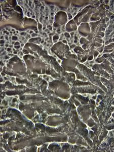
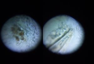
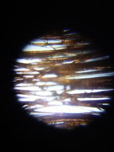
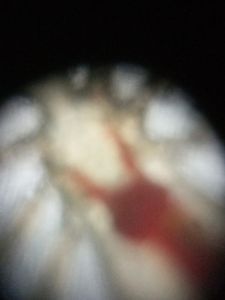
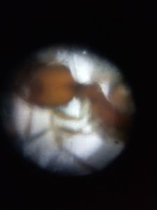
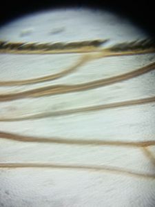
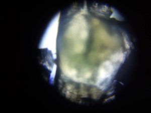
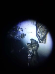
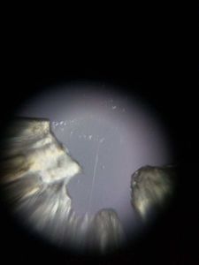
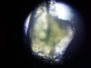
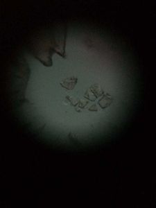
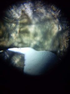
Micro Image on Eye discharge: “Sleep”, or rheum in your eyes
 Jun 03, 2020 • 10:55 PM UTC
 Unknown Location
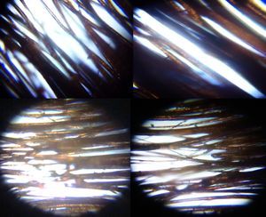
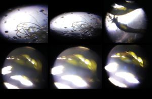
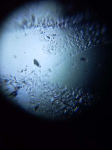
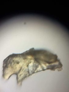
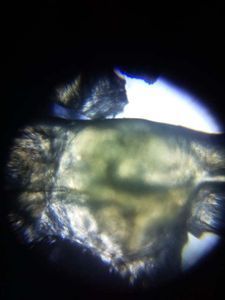
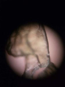
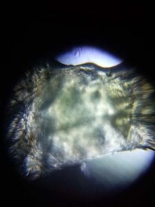
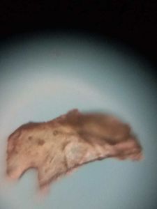
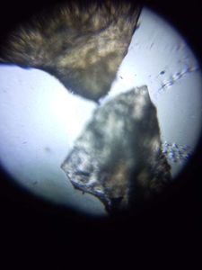
 140x Magnification
 Unknown
Dr. Vemuri SRS Praveen Kumar
I, Dr. Vemuri .S.R.S Praveen Kumar from a reputed family in the Ongole, Praksasm District, Andhra Pradesh in India. My father is a gold Appraiser in Andhra bank. I am a Post Graduate of Master of Technology (M.Tech) from the prestigious University of Tamilnadu, Shanmuga Arts, Science, Technology and Research Academy (SASTRA). I have been an Academically Diligent student throughout my high school & college, having scored 82.0% in X, 85.30% in XII and an aggregate of 6.89 (CGPA) in my B.Tech & 7.37 (CGPA) in my M.Tech. Area of Research Interest – My area of research interest includes Nanotechnology and its applications, Material Science and Engineering, Nano structured Materials, Optical Filters, Rugate, Graded designs and Thin Films coatings. Research Background – Till now I have worked in the field of synthesis of nano particles like ZnO Nano Rods preparation by Solution combustion method. I have also worked in the field of Thin films coating Techniques in SCL, Mohali in the device of CMUT (capacitive Micro machined Ultrasonic Transducer) for ultra sound imaging application. I have studied spectroscopic techniques in detail in my coursework and I have experience of handling Spray Pyrolysis Technique, AFM, spin coater, Electron beam Coating unit etc. Future Research Interest- In the future I would like to continue doing work in the field of nanostructures, and thin film coatings, for futuristic applications and study their mechanical, magnetic, and optical properties. In addition to this, I would like to combine the interdisciplinary approach of various fields in my work. “The Way to get started is to quit talking and begin doing” – Walt Disney
I wake early morning and find that there is yellow eyes mucus pus formed on edge of my eyes, immediately I collected the samples and open my foldscope kit and interested to see its microscopic images, and its usages and why its generated from eyes.
This post gives small information on this concept of eyes pus and its details.

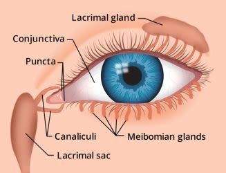
Eye discharge, or “sleep” in your eyes, is a combination of mucus, oil, skin cells and other debris that accumulates in the corner of your eye while you sleep. It can be wet and sticky or dry and crusty, depending on how much of the liquid in the discharge has evaporated.
Sometimes called rheum, eye discharge has a protective function, removing waste products and potentially harmful debris from the tear film and the front surface of your eyes.
Your eyes produce mucus throughout the day, but a continuous thin film of tears bathes your eyes when you blink, flushing out the rheum before it hardens in your eyes.
When you’re asleep — and not blinking — eye discharge collects and crusts in the corners of your eyes and sometimes along the lash line, hence the term “sleep in your eyes.”
Some sleep in your eyes upon waking is normal, but excessive eye discharge, especially if it’s green or yellow in color and accompanied by blurry vision , light sensitivity or eye pain , can indicate a serious eye infection or eye disease and should be promptly examined by your eye doctor.
The remaining information can be seen from the site :
https://www.allaboutvision.com/en-in/conditions/eye-discharge/#:~:text=Also%20called%20MGD%2C%20this%20malfunctioning,by%20an%20infected%20eyelash%20follicle.

 0 Applause
 0 Comments