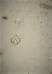
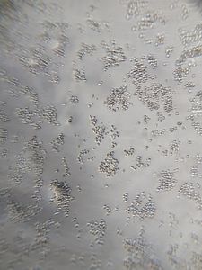
Differential Interference Contrast microscopy(DIC)
 Aug 27, 2020 • 8:37 AM UTC
 Unknown Location
 140x Magnification
 Unknown
Anik Khan
Learn about the author...
5posts
4comments
1locations
For some days I’ve been thinking about this type of microscopy .I saw this on a video on the youtube channel called ‘journey to the microcosmos’,this video blew my mind ,then i remembered that i do have a foldscope,from then i’ve tried to think on how i can do this ,but it was worth the time . .This type of microscopy has special type of 3D effect which is most fascinating .This is mainly about some fascinating optics which I’ve done some research on the internet(Since I’m a physics lover).
HOW DOES DIC WORK?
Light emitted from the source is linearly polarised by passing through a polariser. The linearly polarised beam of light enters an objective-specific prism, which splits it into two rays that vibrate perpendicular to each other. The rays are parallel as they pass through a condenser, but as they are vibrating perpendicular to each other, they are unable to cause interference.
The split beams pass through the specimen. The specimen’s varying thickness and refractive indices alter the wave paths of the beams. They then enter the objective, where they are focussed above the rear focal plane. The two beams enter a second prism, in the nosepiece, which combines them. Because the beams passed through different parts of the specimen, they have different lengths.
The analyser, which is a second polariser, brings the vibrations of the beams into the same plane and axis, causing destructive and constructive interference to occur between the two wavefronts. The light then travels to the eyepiece or camera, where a DIC image with differences in intensity and colour, can be seen.
please think about this type of microscopy (anyone down in the comments) and how we implement this on foldscope ,this could be a change on how we think about the foldscope.
I’m also attaching the link of the video i’ve mentioned above, below
Sign in to commentNobody has commented yet... Share your thoughts with the author and start the discussion!

 0 Applause
 0 Comments