Observation of Onion Cells under a Foldscope
 Nov 02, 2020 • 6:25 AM UTC
 Unknown Location
 140x Magnification
 Microorganisms
Manas Singh Bhati
Learn about the author...
1posts
0comments
1locations
View in Media Gallery
Hi everyone, I’m Manas, a 11th grader from Bangalore, India. I am particularly interested in studying the phenomena of patterning of flower petals using the foldscope.
Before analysing petal patterning, I wanted to work on a simple experiment with onion cells to help me understand the workings of a foldscope.
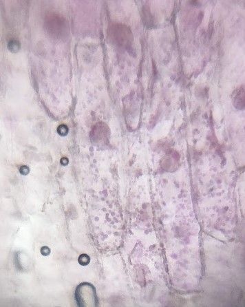
I peeled a few samples of onion cells from the same layer. I soaked the samples of onion cells in a beaker containing RO-filtered water for 30 minutes.
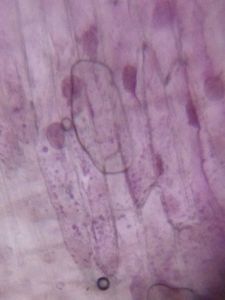
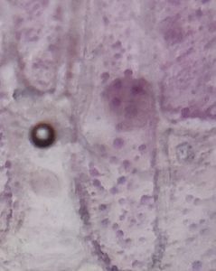
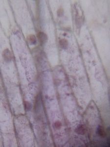
The images of the onion cells that I captured are shown below:
Before analysing petal patterning, I wanted to work on a simple experiment with onion cells to help me understand the workings of a foldscope.
I peeled a few samples of onion cells from the same layer. I soaked the samples of onion cells in a beaker containing RO-filtered water for 30 minutes.
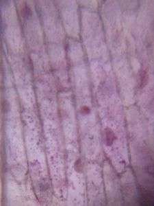
The images of the onion cells that I captured are shown below:


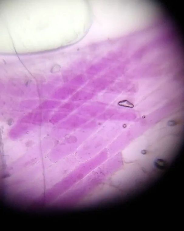
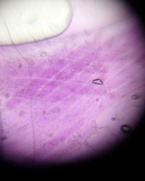
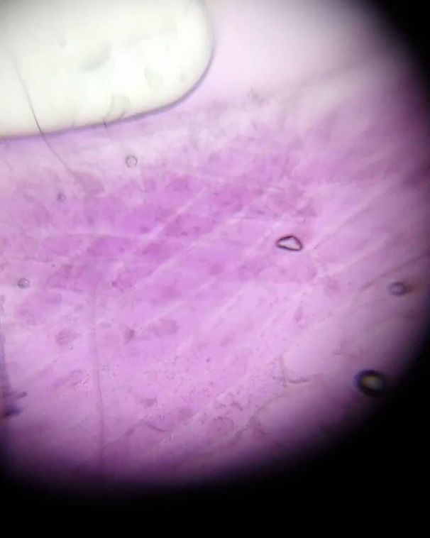
A large purple vacuole can be clearly observed, along with a faint nucleus in some pictures. I took this experiment further by soaking onions in a saturated salt solution. My goal was to understand the effect of osmosis on the microscopy pictures. Attached is the video of these cells while osmosis was taking place (32x):
View in Media Gallery
Also attached are the images from the same video at different times:
View in Media Gallery
0 s (32x)
View in Media Gallery
30 s (32x)
View in Media Gallery
63 s (32x) As time progresses, the extracellular solution is seen to become more and more purple, making the cells paler, as expected.
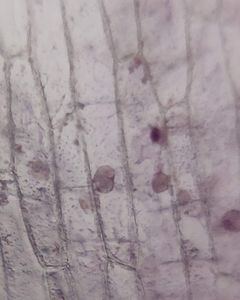
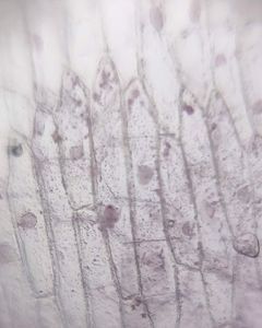
Here are some pictures I took of other onion cells. This is not a part of the experiment, I just captured these images for fun!
Here are some pictures I took of other onion cells. This is not a part of the experiment, I just captured these images for fun!


This is my first experiment with the foldscope. I would be posting more content in the near future on patterning of flowers, using the foldscope.
Signing off,
Manas.
Signing off,
Manas.
Sign in to commentNobody has commented yet... Share your thoughts with the author and start the discussion!
More Posts from Manas Singh Bhati
No more posts from this author.