Soil, Water, Plant, Fruit!
 Nov 03, 2020 • 3:07 PM UTC
 Unknown Location
 140x Magnification
 Unknown
Bryan To
Learn about the author...
1posts
0comments
1locations
View in Media Gallery
Hello! Bryan To, here! This is my first ever Foldscope blog, and I’m really excited.
First off, I must express how amazed I am by Foldscope. Every time I watch Manu Prakash’s Ted Talk, there are shivers down my spine and goosebumps everywhere. It truly is an amazing myriad of feats: they reduced thousands of dollars to tiny cents and allowed the miraculous images of microbial visualization to be accessible to all. Scientists, civilians, teachers, students, KIDS. I remember in high school when I first looked into a microscope and saw microbial movement. It was mind-blowing. To see these foldscopes distributed everywhere – that allows kids to be exposed early to the intricate and invisible structures around them, to be exposed to science in such an amazing, accessible, and cheap fashion – it truly makes me smile.
I felt like a kid again when my class received our foldscopes. I’m a junior in college. Did using electron microscopes make my jaw drop? Well yes, but that’s not the point. When I held the fully built piece of paper in my hands, I marveled at the foldscope like an amazing toy. I was so excited and wished to test it on EVERYTHING.
Immediately I was like, DAD, CAN I USE YOUR DIABETES BLOOD NEEDLE THINGIE? I WANNA SEE MY BLOOD UNDER A PAPER MICROSCOPE . Luckily, my parents with common sense stopped me from going too far.
My parents were in their 50s and 60s and refugees from Vietnam. My dad never finished his high school education. I showed them the Ted Talks, the Youtube videos, everything. We all marveled together at the transformation from a light microscope to a piece of paper. I was so happy to be able to show them the foldscope and let them see through a microscope. They got to experience a part of the career I want to pursue!
Actual Blog Content
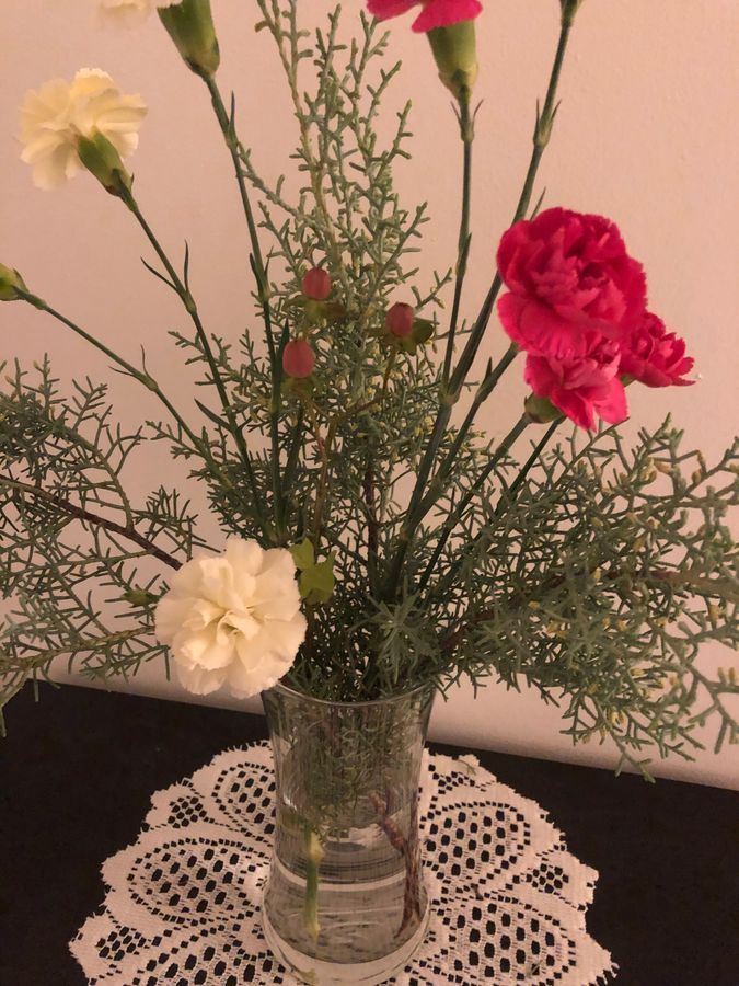
So today (finally xD), I am posting a blog about a comparison across the different structures of household plant mediums. My mom keeps some flowers around the house, some in soil, some in water, so I was curious about what soil versus water with plants in them looked like under a microscope.
My first observation involves the Ghost-Plant, graptopetalum paraguayense, a succulent plant of the jade plant family.
First off, I must express how amazed I am by Foldscope. Every time I watch Manu Prakash’s Ted Talk, there are shivers down my spine and goosebumps everywhere. It truly is an amazing myriad of feats: they reduced thousands of dollars to tiny cents and allowed the miraculous images of microbial visualization to be accessible to all. Scientists, civilians, teachers, students, KIDS. I remember in high school when I first looked into a microscope and saw microbial movement. It was mind-blowing. To see these foldscopes distributed everywhere – that allows kids to be exposed early to the intricate and invisible structures around them, to be exposed to science in such an amazing, accessible, and cheap fashion – it truly makes me smile.
I felt like a kid again when my class received our foldscopes. I’m a junior in college. Did using electron microscopes make my jaw drop? Well yes, but that’s not the point. When I held the fully built piece of paper in my hands, I marveled at the foldscope like an amazing toy. I was so excited and wished to test it on EVERYTHING.
Immediately I was like, DAD, CAN I USE YOUR DIABETES BLOOD NEEDLE THINGIE? I WANNA SEE MY BLOOD UNDER A PAPER MICROSCOPE . Luckily, my parents with common sense stopped me from going too far.
My parents were in their 50s and 60s and refugees from Vietnam. My dad never finished his high school education. I showed them the Ted Talks, the Youtube videos, everything. We all marveled together at the transformation from a light microscope to a piece of paper. I was so happy to be able to show them the foldscope and let them see through a microscope. They got to experience a part of the career I want to pursue!
Actual Blog Content
So today (finally xD), I am posting a blog about a comparison across the different structures of household plant mediums. My mom keeps some flowers around the house, some in soil, some in water, so I was curious about what soil versus water with plants in them looked like under a microscope.
My first observation involves the Ghost-Plant, graptopetalum paraguayense, a succulent plant of the jade plant family.

View in Media Gallery
Ghost-Plant
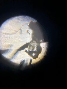
graptopetalum paraguayense Here’s a video recorded by my phone looking through the foldscope of a small part of the soil.
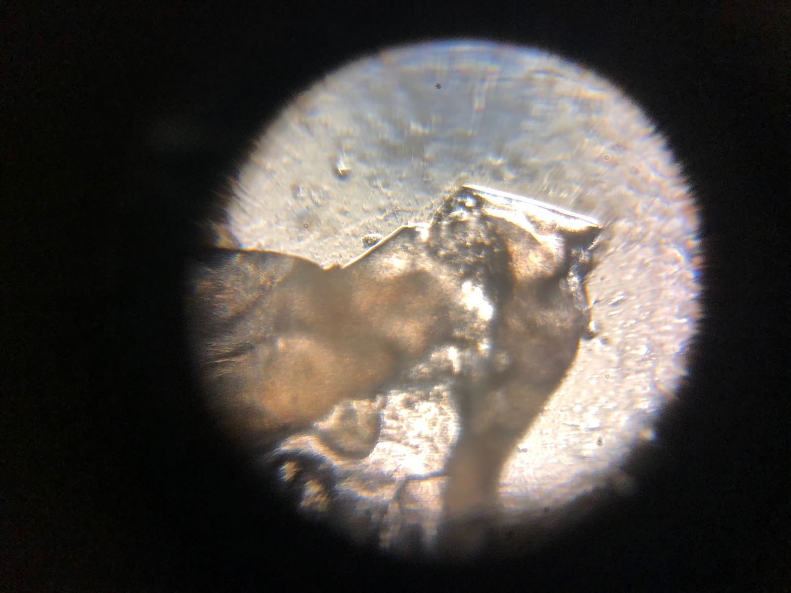
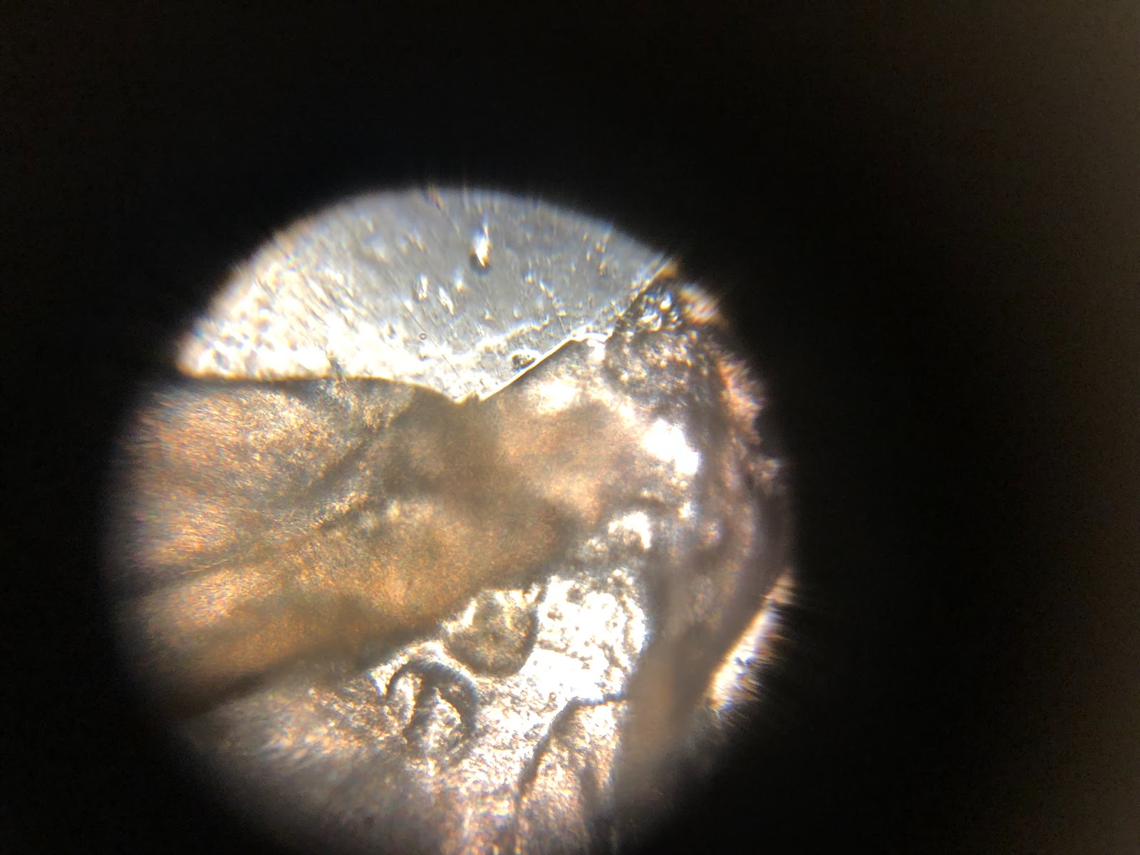
Soil + Ghost Plant
graptopetalum paraguayense Here’s a video recorded by my phone looking through the foldscope of a small part of the soil.
Soil + Ghost Plant
You can see the soil is very dense black / brown under the microscope. It’s super clumped together. Here, I was focusing on a translucent bulbous shape that had spots of green. Maybe the chloroplast? Here’s a still picture (see video above for more.)

So cool!
Moving from soil to a water medium, I was curious about water that contained my mom’s Moss-rose Purslane, Portulaca grandiflora, a flowering plant introduced from its native region of Argentina, southern Brazil, Uruguay.
Moving from soil to a water medium, I was curious about water that contained my mom’s Moss-rose Purslane, Portulaca grandiflora, a flowering plant introduced from its native region of Argentina, southern Brazil, Uruguay.
View in Media Gallery
Moss-rose Purslane
Portulaca grandiflora
Here’s the water video.
Portulaca grandiflora
Here’s the water video.
Comparing it to past experiences of using light microscopes to see pond or dirty water, this water was much cleaner. Still, it’s full of things which I presume are small plant matter or other organisms. Or dirt / small matter. Here I focus on one interesting thing that reminded me of a heart. I don’t know why, but my thought was a heart when I saw it (see video for the angle – what do you guys think?) My brother says that from the photo below, it was more like a crab. Honestly, it’s like looking at a cloud and interpreting differently depending on the person. I’m not even sure if this was an organism or just a dirt speck.

View in Media Gallery
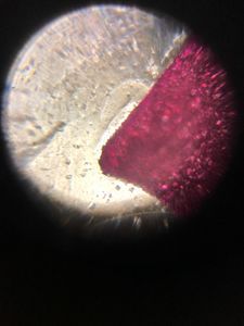
Next, here’s the flower petal of the moss-rose purslane. Look at this specific petal’s striking red color!
Flower Petal
Flower Petal
By the way, all vibrating sound in the background is my phone not playing well with the magnet and reacting weirdly. But as for the petal, it was a beautiful red hue. I specifically looked at the border, where it was like rubies lining the edge of the petal. It looks like a different mars landscape almost!
Flower Petal pt 2
Flower Petal pt 2


Absolutely spectacular. If this was what it looked liked human-sized, I would’ve thought it was scales or a metal (back to the ruby similarity).
Finally, we have a grapefruit. Just for fun. I wanted to compare a fruit with a flowering plant.
Grapefruit!
Finally, we have a grapefruit. Just for fun. I wanted to compare a fruit with a flowering plant.
Grapefruit!
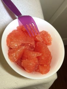

This was not as clear red color as the petal under the microscope. It was more light translucent pink, but that’s because I stretched it thin so it wouldn’t block the light. You can see the cell wall! Under a microscope, it looked more like slime than anything else.
Plant cell walls have always been cool to me – plant physiology of wall structure finds many important functions such as providing structure and shape to even intercellular communication in things like defense responses. I wonder about the tradeoffs in plant cell walls. Sturdyness versus flexibility. How much stress tolerance can cell walls take – does it give in and thus incurs changes in cell wall composition? A lot of curiosities!
Thanks for reading!
Bryan To
I conducted this project as part of Professor Pringle’s EEB321 class at Princeton University.
Sign in to commentNobody has commented yet... Share your thoughts with the author and start the discussion!
More Posts from Bryan To
No more posts from this author.