Skin cells: from cheek to dust
 Nov 05, 2020 • 11:29 AM UTC
 Unknown Location
 140x Magnification
 Unknown
Leah Smith
Learn about the author...
1posts
0comments
1locations

View in Media Gallery
I conducted this project as part of Professor Pringle’s EEB321 class at Princeton University.
After assembling my foldscope, which took my unsteady hands and “I don’t follow directions brain” a minute, I promptly started observing the world around me.
I tried clothing fuzz, cheek cells, and foot skin. (I know, a little bit gross) but I am fascinated by the human body and the very different cell types and formations.
While cheek cells and epidermis layer of feet cells are cool, I was curious about the ecological community of skin in general. I knew that the inner layers of skin have more blood flow, like the cheek cells, being the dermis and the hypodermis. I knew that the outer layers of skin, like what was on my foot, would be the epidermis. And I knew that what would be on my dresser or under my bed would be most likely flaked off skin cells. And so I set out to observe the differences.
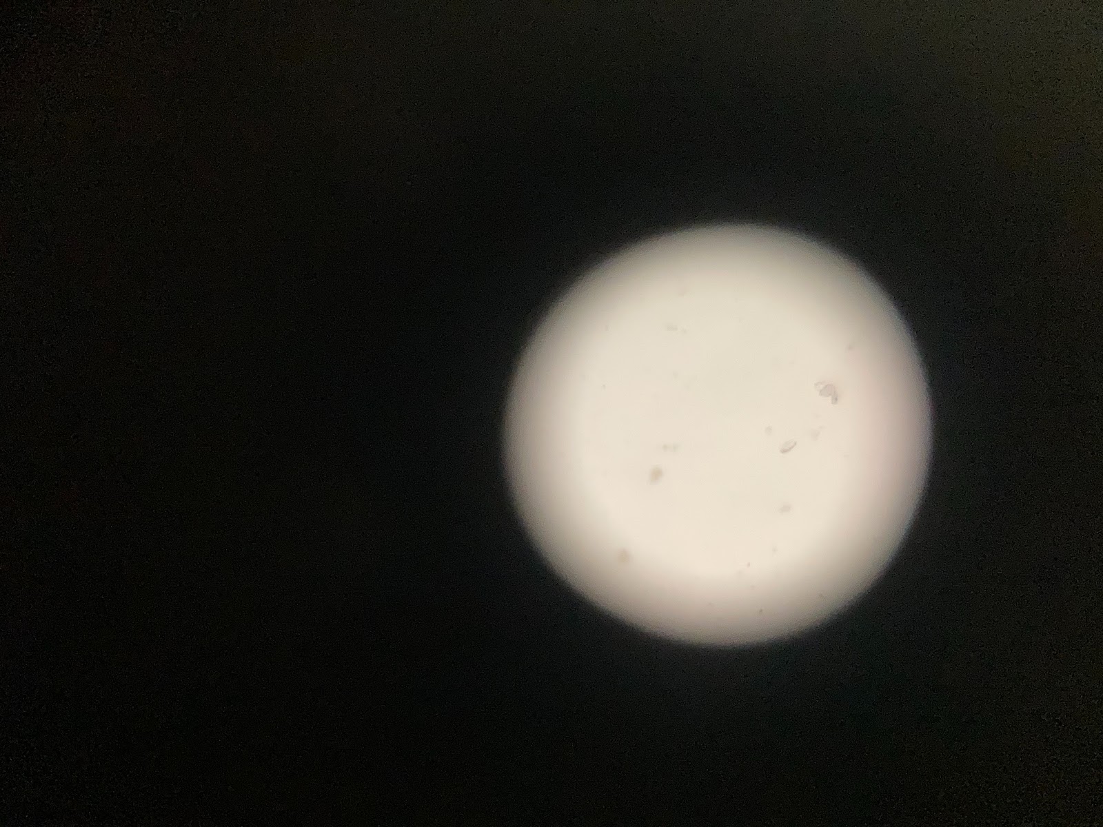
Here we can see my cheek cells. Small and cocci shaped they were pretty spread out. Probably because of the salvia that was involved.
After assembling my foldscope, which took my unsteady hands and “I don’t follow directions brain” a minute, I promptly started observing the world around me.
I tried clothing fuzz, cheek cells, and foot skin. (I know, a little bit gross) but I am fascinated by the human body and the very different cell types and formations.
While cheek cells and epidermis layer of feet cells are cool, I was curious about the ecological community of skin in general. I knew that the inner layers of skin have more blood flow, like the cheek cells, being the dermis and the hypodermis. I knew that the outer layers of skin, like what was on my foot, would be the epidermis. And I knew that what would be on my dresser or under my bed would be most likely flaked off skin cells. And so I set out to observe the differences.
Here we can see my cheek cells. Small and cocci shaped they were pretty spread out. Probably because of the salvia that was involved.


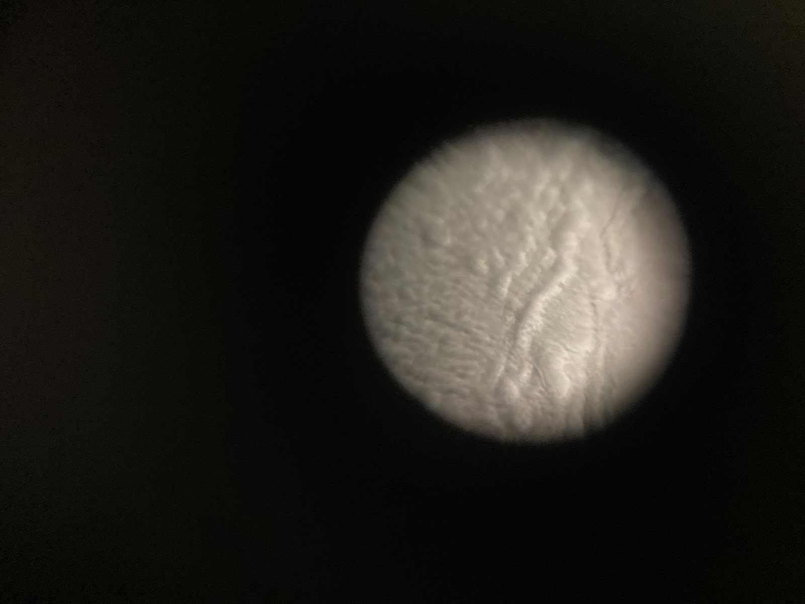
Here is a small sample of the callus of my foot!
More rigid, there seems to be more lines and structure to these cells, like little blocks.
More rigid, there seems to be more lines and structure to these cells, like little blocks.


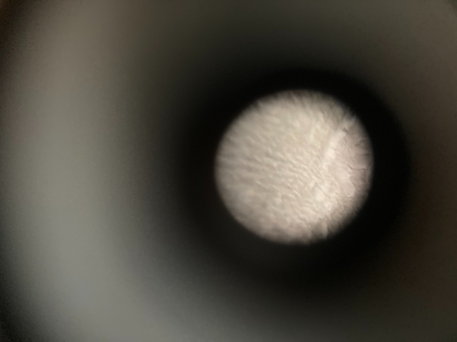
Here is dead skin tissue. When I zoom in it has many more lines and fissures than the foot cells. Probably because it is dead skin from my entire body and not just my feet!
View in Media Gallery
The difference in cell types is incredibly fascinating! I love how even the shape and structure and size of the cell changes depending on where it was found (cheek vs foot)
I also loved looking at the differences between dead skin still attached to my body, and dead skin on the ground in dust form. Also, note that the prevalence of dead skin in the dust is interrupted by dust mite products as well. It didn’t capture well on the foldscope since dust is so big, but the big blocky substituents on the right of the photo may be termite droppings.
I also loved looking at the differences between dead skin still attached to my body, and dead skin on the ground in dust form. Also, note that the prevalence of dead skin in the dust is interrupted by dust mite products as well. It didn’t capture well on the foldscope since dust is so big, but the big blocky substituents on the right of the photo may be termite droppings.
Sign in to commentNobody has commented yet... Share your thoughts with the author and start the discussion!
More Posts from Leah Smith
No more posts from this author.